Application
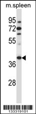
TBCC Antibody (N-term ) western blot analysis in mouse spleen tissue lysates (35ug/lane).This demonstrates the TBCC antibody detected the TBCC protein (arrow).| Product Name | TBCC Antibody (N-term ) |
|---|---|
| Antibody Type | Primary Antibodies |
| Antigen Alias | TBCC; Tubulin-specific chaperone C; Tubulin-folding cofactor C |
| Clonality | Polyclonal |
|---|---|
| Isotype | Ig |
| Host Species | Rabbit |
| Tested Applications | WB |
| WB:1:100~500 |
|
| Species Reactivity | Human |
| Concentration | 1mg/ml |
| Alternative Names | TBCC Tubulin-specific chaperone C Tubulin-folding cofactor C |
|---|---|
| Molecular Weight(MW) | 39248 Da |
| Function | Tubulin-folding protein; involved in the final step of the tubulin folding pathway |
| Tissue Specificity | Expressed in the retina. Expressed in the rod and cone photoreceptors, extending from the inner segments (IS), through the outer nuclear layer (ONL) and into the synapses in the outer plexiform layer (OPL). Strongly expressed to the photoreceptor connecting cilium at the tips of the IS (at protein level) |
| Cellular Localization | Cytoplasm. Note=Detected predominantly in the photoreceptor connecting cilium |
| Entrez Gene | 6903 |
|---|---|
| Nucleotide Accession | NP_003183.1 |
Application
TBCC Antibody (N-term ) western blot analysis in mouse spleen tissue lysates (35ug/lane).This demonstrates the TBCC antibody detected the TBCC protein (arrow).| Application Notes | WB:1:100~500 |
|---|
| Form | Liquid |
|---|---|
| Storage Instructions | For short-term storage, store at 4° C. For long-term storage, aliquot and store at -20ºC or below. Avoid multiple freeze-thaw cycles. |
| Storage Buffer | Purified polyclonal antibody supplied in PBS with 0.09% (W/V) sodium azide. This antibody is purified through a protein A column, followed by peptide affinity purification. |


