Application
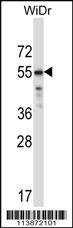
TFCP2L1 Antibody (Center) western blot analysis in WiDr cell line lysates (35ug/lane).This demonstrates the TFCP2L1 antibody detected the TFCP2L1 protein (arrow).Application
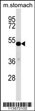
TFCP2L1 Antibody (Center) western blot analysis in mouse stomach tissue lysates (35ug/lane).This demonstrates the TFCP2L1 antibody detected the TFCP2L1 protein (arrow).Application
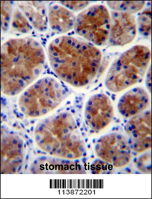
TFCP2L1 Antibody (Center) immunohistochemistry analysis in formalin fixed and paraffin embedded human stomach tissue followed by peroxidase conjugation of the secondary antibody and DAB staining.This data demonstrates the use of TFCP2L1 Antibody (Center) for immunohistochemistry. Clinical relevance has not been evaluated.Application
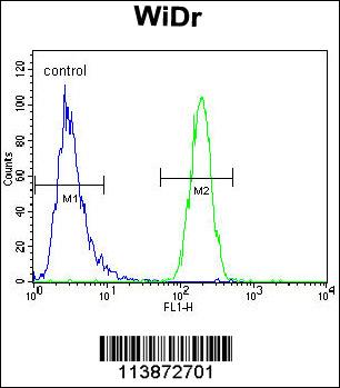
TFCP2L1 Antibody (Center) flow cytometric analysis of WiDr cells (right histogram) compared to a negative control cell (left histogram).FITC-conjugated goat-anti-rabbit secondary antibodies were used for the analysis.| Product Name | TFCP2L1 Antibody (Center) |
|---|---|
| Antibody Type | Primary Antibodies |
| Clonality | Polyclonal |
|---|---|
| Isotype | Ig |
| Host Species | Rabbit |
| Tested Applications | WBIHCFC |
| WB:1:100~500 IHC |
|
| Species Reactivity | Human |
| Concentration | 1mg/ml |
| Gene Synonyms | CRTR1, LBP9 |
|---|---|
| Alternative Names | TFCP2L1 CRTR1 LBP9 Transcription factor CP2-like protein 1 CP2-related transcriptional repressor 1 Transcription factor LBP-9 |
| Molecular Weight(MW) | 54627 Da |
| Function | Transcriptional suppressor. May suppress UBP1-mediated transcriptional activation. Required for normal duct development in the salivary gland and kidney (By similarity). Modulates the placental expression of CYP11A1 |
| Tissue Specificity | Highly expressed in placental JEG-3 cells and very low levels of expression in non-steroidogenic cells. No expression was seen in adrenal NCI-H295A cells or in adrenal tissue |
| Cellular Localization | Nucleus. |
| Entrez Gene | 29842 |
|---|---|
| Nucleotide Accession | NP_055368.1 |
Application
TFCP2L1 Antibody (Center) western blot analysis in WiDr cell line lysates (35ug/lane).This demonstrates the TFCP2L1 antibody detected the TFCP2L1 protein (arrow).
Application
TFCP2L1 Antibody (Center) western blot analysis in mouse stomach tissue lysates (35ug/lane).This demonstrates the TFCP2L1 antibody detected the TFCP2L1 protein (arrow).
Application
TFCP2L1 Antibody (Center) immunohistochemistry analysis in formalin fixed and paraffin embedded human stomach tissue followed by peroxidase conjugation of the secondary antibody and DAB staining.This data demonstrates the use of TFCP2L1 Antibody (Center) for immunohistochemistry. Clinical relevance has not been evaluated.
Application
TFCP2L1 Antibody (Center) flow cytometric analysis of WiDr cells (right histogram) compared to a negative control cell (left histogram).FITC-conjugated goat-anti-rabbit secondary antibodies were used for the analysis.| Application Notes | WB:1:100~500 IHC |
|---|
| Form | Liquid |
|---|---|
| Storage Instructions | For short-term storage, store at 4° C. For long-term storage, aliquot and store at -20ºC or below. Avoid multiple freeze-thaw cycles. |
| Storage Buffer | Purified polyclonal antibody supplied in PBS with 0.09% (W/V) sodium azide. This antibody is purified through a protein A column, followed by peptide affinity purification. |


