Application
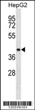
TID1 Antibody (N-term) western blot analysis in HepG2 cell line lysates (35ug/lane).This demonstrates the TID1 antibody detected the TID1 protein (arrow).| Product Name | TID1 Antibody (N-term) |
|---|---|
| Antibody Type | Primary Antibodies |
| Clonality | Polyclonal |
|---|---|
| Isotype | Ig |
| Host Species | Rabbit |
| Tested Applications | WB |
| WB:1:100~500 |
|
| Species Reactivity | Human |
| Concentration | 1mg/ml |
| Gene Synonyms | HCA57, TID1 |
|---|---|
| Alternative Names | DNAJA3 HCA57 TID1 DnaJ homolog subfamily A member 3 mitochondrial DnaJ protein Tid-1 Hepatocellular carcinoma-associated antigen 57 Tumorous imaginal discs protein Tid56 homolog |
| Molecular Weight(MW) | 52489 Da |
| Function | Modulates apoptotic signal transduction or effector structures within the mitochondrial matrix. Affect cytochrome C release from the mitochondria and caspase 3 activation, but not caspase 8 activation. Isoform 1 increases apoptosis triggered by both TNF and the DNA-damaging agent mytomycin C; in sharp contrast, isoform 2 suppresses apoptosis. Can modulate IFN-gamma- mediated transcriptional activity. Isoform 2 may play a role in neuromuscular junction development as an effector of the MUSK signaling pathway |
| Tissue Specificity | Widely expressed with highest levels in heart, liver, lung and skeletal muscles. Also expressed in keratinocytes |
| Cellular Localization | Mitochondrion matrix. Cytoplasm, cytosol (By similarity). Cell junction, synapse, postsynaptic cell membrane; Peripheral membrane protein (By similarity). Note=Recruited to the postsynaptic cell membrane of the neuromuscular junction through interaction with MUSK (By similarity) |
| Entrez Gene | 9093 |
|---|---|
| Nucleotide Accession | NP_005138.3 NP_001128582.1 |
Application
TID1 Antibody (N-term) western blot analysis in HepG2 cell line lysates (35ug/lane).This demonstrates the TID1 antibody detected the TID1 protein (arrow).| Application Notes | WB:1:100~500 |
|---|
| Form | Liquid |
|---|---|
| Storage Instructions | For short-term storage, store at 4° C. For long-term storage, aliquot and store at -20ºC or below. Avoid multiple freeze-thaw cycles. |
| Storage Buffer | Purified polyclonal antibody supplied in PBS with 0.09% (W/V) sodium azide. This antibody is purified through a protein A column, followed by peptide affinity purification. |


