Application
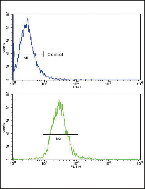
Flow cytometric analysis of NCI-H460 cells using TLR6 Antibody (C-term) (bottom histogram) compared to a negative control cell (top histogram). FITC-conjugated goat-anti-rabbit secondary antibodies were used for the analysis.| Product Name | TLR6 Antibody (C-term) |
|---|---|
| Antibody Type | Primary Antibodies |
| Clonality | Polyclonal |
|---|---|
| Isotype | Ig |
| Host Species | Rabbit |
| Tested Applications | FC |
| FC:1:10~50 |
|
| Species Reactivity | Human |
| Concentration | 1mg/ml |
| Alternative Names | TLR6 Toll-like receptor 6 CD_antigen=CD286 Flags: Precursor |
|---|---|
| Molecular Weight(MW) | 91880 Da |
| Function | Participates in the innate immune response to Gram- positive bacteria and fungi. Specifically recognizes diacylated and, to a lesser extent, triacylated lipopeptides. Acts via MYD88 and TRAF6, leading to NF-kappa-B activation, cytokine secretion and the inflammatory response. Recognizes mycoplasmal macrophage- activating lipopeptide-2kD (MALP-2), soluble tuberculosis factor (STF), phenol-soluble modulin (PSM) and B.burgdorferi outer surface protein A lipoprotein (OspA-L) cooperatively with TLR2 |
| Tissue Specificity | Detected in monocytes, CD11c+ immature dendritic cells, plasmacytoid pre-dendritic cells and dermal microvessel endothelial cells |
| Cellular Localization | Cell membrane; Single-pass type I membrane protein (By similarity). Cytoplasmic vesicle, phagosome membrane; Single-pass type I membrane protein (By similarity) |
| Entrez Gene | 10333 |
|---|
Application
Flow cytometric analysis of NCI-H460 cells using TLR6 Antibody (C-term) (bottom histogram) compared to a negative control cell (top histogram). FITC-conjugated goat-anti-rabbit secondary antibodies were used for the analysis.| Application Notes | FC:1:10~50 |
|---|
| Form | Liquid |
|---|---|
| Storage Instructions | For short-term storage, store at 4° C. For long-term storage, aliquot and store at -20ºC or below. Avoid multiple freeze-thaw cycles. |
| Storage Buffer | Purified polyclonal antibody supplied in PBS with 0.09% (W/V) sodium azide. This antibody is purified through a protein G column, eluted with high and low pH buffers and neutralized immediately, followed by dialysis against PBS. |


