Application
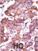
Formalin-fixed and paraffin-embedded human cancer tissue reacted with the primary antibody, which was peroxidase-conjugated to the secondary antibody, followed by AEC staining. This data demonstrates the use of this antibody for immunohistochemistry; clinical relevance has not been evaluated. BC = breast carcinoma; HC = hepatocarcinoma.Application
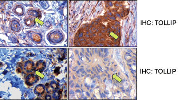
Middle panels, TOLLIP expression in matched normal and tumor breast samples. Here the TOLLIP stain in the invasive ductal carcinoma cells shows a much stronger signal compared with the normal ductal cells. Bottom panels, TOLLIP expression in matched normal and tumor breast samples. In this case, the signal for TOLLIP in the normal ductal cells is stronger than that in the invasive ductal carcinoma cells.Application
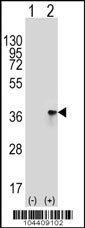
Western blot analysis of TOLLIP (arrow) using rabbit polyclonal TOLLIP Antibody (A236) . 293 cell lysates (2 ug/lane) either nontransfected (Lane 1) or transiently transfected (Lane 2) with the TOLLIP gene.| Product Name | TOLLIP Antibody (C-term) |
|---|---|
| Antibody Type | Primary Antibodies |
| Antigen Alias | TOLLIP; Toll-interacting protein |
| Clonality | Polyclonal |
|---|---|
| Isotype | Ig |
| Host Species | Rabbit |
| Tested Applications | IHCWB |
| IHC:1:50~100 WB |
|
| Species Reactivity | Human |
| Concentration | 1mg/ml |
| Alternative Names | TOLLIP Toll-interacting protein |
|---|---|
| Molecular Weight(MW) | 30282 Da |
| Function | Component of the signaling pathway of IL-1 and Toll-like receptors. Inhibits cell activation by microbial products Recruits IRAK1 to the IL-1 receptor complex. Inhibits IRAK1 phosphorylation and kinase activity |
| Tissue Specificity | This TOLLIP antibody is generated from rabbits immunized with a KLH conjugated synthetic peptide between 228~258 amino acids from the C-terminal region of human TOLLIP. |
| Cellular Localization | Cytoplasm (Probable). |
| Entrez Gene | 54472 |
|---|
Application
Formalin-fixed and paraffin-embedded human cancer tissue reacted with the primary antibody, which was peroxidase-conjugated to the secondary antibody, followed by AEC staining. This data demonstrates the use of this antibody for immunohistochemistry; clinical relevance has not been evaluated. BC = breast carcinoma; HC = hepatocarcinoma.
Application
Middle panels, TOLLIP expression in matched normal and tumor breast samples. Here the TOLLIP stain in the invasive ductal carcinoma cells shows a much stronger signal compared with the normal ductal cells. Bottom panels, TOLLIP expression in matched normal and tumor breast samples. In this case, the signal for TOLLIP in the normal ductal cells is stronger than that in the invasive ductal carcinoma cells.
Application
Western blot analysis of TOLLIP (arrow) using rabbit polyclonal TOLLIP Antibody (A236) . 293 cell lysates (2 ug/lane) either nontransfected (Lane 1) or transiently transfected (Lane 2) with the TOLLIP gene.| Application Notes | IHC:1:50~100 WB |
|---|
| Form | Liquid |
|---|---|
| Storage Instructions | For short-term storage, store at 4° C. For long-term storage, aliquot and store at -20ºC or below. Avoid multiple freeze-thaw cycles. |
| Storage Buffer | Purified polyclonal antibody supplied in PBS with 0.09% (W/V) sodium azide. This antibody is purified through a protein G column, eluted with high and low pH buffers and neutralized immediately, followed by dialysis against PBS. |


