Application
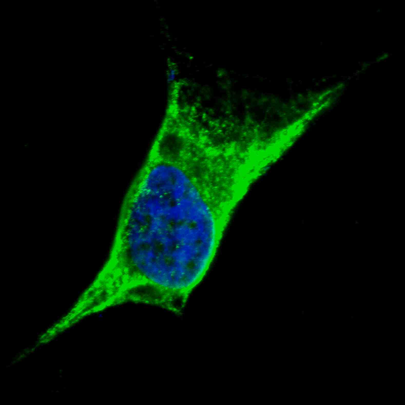
Fluorescent confocal image of SY5Y cells stained with Vimentin (C-term) antibody. SY5Y cells were fixed with 4% PFA (20 min), permeabilized with Triton X-100 (0.2%, 30 min). Cells were then incubated with AP2739b Vimentin (C-term) primary antibody (1:100, 2 h at room temperature). For secondary antibody, Alexa Fluor® 488 conjugated donkey anti-rabbit antibody (green) was used (1:1000, 1h). Nuclei were counterstained with Hoechst 33342 (blue) (10 μg/ml, 5 min). Note the highly specific localization of the Vimentin immunosignal to the cytoskeleton, supported by Human Protein Atlas Data (http://www.proteinatlas.org/ENSG00000026025).Application
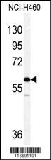
Western blot analysis of Vimentin antibody (C-term) in NCI-H460 cell line lysates (35ug/lane). Vimentin (arrow) was detected using the purified Pab .Application
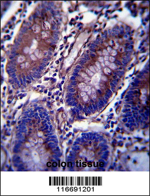
Vimentin Antibody (C-term) immunohistochemistry analysis in formalin fixed and paraffin embedded human colon tissue followed by peroxidase conjugation of the secondary antibody and DAB staining.This data demonstrates the use of Vimentin Antibody (C-term) for immunohistochemistry. Clinical relevance has not been evaluated.Application
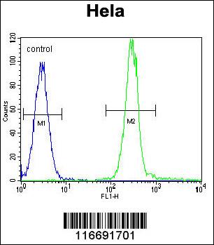
Vimentin Antibody (C-term) flow cytometric analysis of Hela cells (right histogram) compared to a negative control cell (left histogram).FITC-conjugated goat-anti-rabbit secondary antibodies were used for the analysis.| Product Name | Vimentin Antibody (C-term) |
|---|---|
| Antibody Type | Primary Antibodies |
| Clonality | Polyclonal |
|---|---|
| Isotype | Ig |
| Host Species | Rabbit |
| Tested Applications | IFWBIHCFC |
| IF:1:100 WB |
|
| Species Reactivity | Human |
| Concentration | 1mg/ml |
| Alternative Names | VIM Vimentin |
|---|---|
| Molecular Weight(MW) | 53652 Da |
| Function | Vimentins are class-III intermediate filaments found in various non-epithelial cells, especially mesenchymal cells Vimentin is attached to the nucleus, endoplasmic reticulum, and mitochondria, either laterally or terminally |
| Tissue Specificity | Highly expressed in fibroblasts, some expression in T- and B-lymphocytes, and little or no expression in Burkitt's lymphoma cell lines. Expressed in many hormone- independent mammary carcinoma cell lines |
| Cellular Localization | Cytoplasm. |
| Entrez Gene | 7431 |
|---|
Application
Fluorescent confocal image of SY5Y cells stained with Vimentin (C-term) antibody. SY5Y cells were fixed with 4% PFA (20 min), permeabilized with Triton X-100 (0.2%, 30 min). Cells were then incubated with AP2739b Vimentin (C-term) primary antibody (1:100, 2 h at room temperature). For secondary antibody, Alexa Fluor® 488 conjugated donkey anti-rabbit antibody (green) was used (1:1000, 1h). Nuclei were counterstained with Hoechst 33342 (blue) (10 μg/ml, 5 min). Note the highly specific localization of the Vimentin immunosignal to the cytoskeleton, supported by Human Protein Atlas Data (http://www.proteinatlas.org/ENSG00000026025).
Application
Western blot analysis of Vimentin antibody (C-term) in NCI-H460 cell line lysates (35ug/lane). Vimentin (arrow) was detected using the purified Pab .
Application
Vimentin Antibody (C-term) immunohistochemistry analysis in formalin fixed and paraffin embedded human colon tissue followed by peroxidase conjugation of the secondary antibody and DAB staining.This data demonstrates the use of Vimentin Antibody (C-term) for immunohistochemistry. Clinical relevance has not been evaluated.
Application
Vimentin Antibody (C-term) flow cytometric analysis of Hela cells (right histogram) compared to a negative control cell (left histogram).FITC-conjugated goat-anti-rabbit secondary antibodies were used for the analysis.| Application Notes | IF:1:100 WB |
|---|
| Form | Liquid |
|---|---|
| Storage Instructions | For short-term storage, store at 4° C. For long-term storage, aliquot and store at -20ºC or below. Avoid multiple freeze-thaw cycles. |
| Storage Buffer | Purified polyclonal antibody supplied in PBS with 0.09% (W/V) sodium azide. This antibody is prepared by Saturated Ammonium Sulfate (SAS) precipitation followed by dialysis against PBS. |


