Application
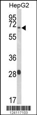
Western blot analysis of WAC Antibody (Center) in HepG2 cell line lysates (35ug/lane). WAC(arrow) was detected using the purified Pab.Application
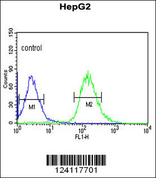
WAC Antibody (Center) flow cytometric analysis of HepG2 cells (right histogram) compared to a negative control cell (left histogram).FITC-conjugated goat-anti-rabbit secondary antibodies were used for the analysis.| Product Name | WAC Antibody (Center) |
|---|---|
| Antibody Type | Primary Antibodies |
| Antigen Alias | WAC; KIAA1844; WW domain-containing adapter protein with coiled-coil |
| Clonality | Polyclonal |
|---|---|
| Isotype | Ig |
| Host Species | Rabbit |
| Tested Applications | WBFC |
| WB:1:100~500 FC |
|
| Species Reactivity | Human |
| Concentration | 1mg/ml |
| Gene Synonyms | KIAA1844 |
|---|---|
| Alternative Names | WAC KIAA1844 WW domain-containing adapter protein with coiled-coil |
| Molecular Weight(MW) | 70724 Da |
| Function | Acts as a linker between gene transcription and histone H2B monoubiquitination at 'Lys-120' (H2BK120ub1). Interacts with the RNA polymerase II transcriptional machinery via its WW domain and with RNF20-RNF40 via its coiled coil region, thereby linking and regulating H2BK120ub1 and gene transcription. Regulates the cell-cycle checkpoint activation in response to DNA damage Positive regulator of amino acid starvation-induced autophagy. May negatively regulate the ubiquitin proteasome pathway |
| Tissue Specificity | This WAC antibody is generated from rabbits immunized with a KLH conjugated synthetic peptide between 291~321 amino acids from the Central region of human WAC. |
| Cellular Localization | Nucleus speckle (By similarity). Nucleus. Note=In distinct nuclear speckles. Colocalizes with pre-mRNA processing complexes (By similarity) |
| Entrez Gene | 51322 |
|---|
Application
Western blot analysis of WAC Antibody (Center) in HepG2 cell line lysates (35ug/lane). WAC(arrow) was detected using the purified Pab.
Application
WAC Antibody (Center) flow cytometric analysis of HepG2 cells (right histogram) compared to a negative control cell (left histogram).FITC-conjugated goat-anti-rabbit secondary antibodies were used for the analysis.| Application Notes | WB:1:100~500 FC |
|---|
| Form | Liquid |
|---|---|
| Storage Instructions | For short-term storage, store at 4° C. For long-term storage, aliquot and store at -20ºC or below. Avoid multiple freeze-thaw cycles. |
| Storage Buffer | Purified polyclonal antibody supplied in PBS with 0.09% (W/V) sodium azide. This antibody is purified through a protein A column, followed by peptide affinity purification. |


