Application
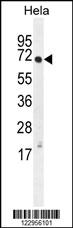
XRCC6 Antibody (Center) western blot analysis in Hela cell line lysates (35ug/lane).This demonstrates the XRCC6 antibody detected the XRCC6 protein (arrow).Application
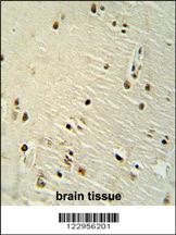
XRCC6 antibody (Center) immunohistochemistry analysis in formalin fixed and paraffin embedded human brain tissue followed by peroxidase conjugation of the secondary antibody and DAB staining. This data demonstrates the use of the XRCC6 antibody (Center) for immunohistochemistry. Clinical relevance has not been evaluated.Application
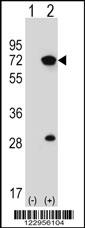
Western blot analysis of XRCC6 (arrow) using rabbit polyclonal XRCC6 Antibody (Center) . 293 cell lysates (2 ug/lane) either nontransfected (Lane 1) or transiently transfected (Lane 2) with the XRCC6 gene.Application
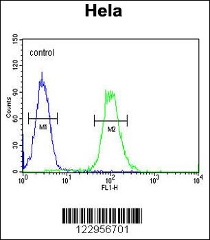
XRCC6 Antibody (Center) flow cytometric analysis of Hela cells (right histogram) compared to a negative control cell (left histogram).FITC-conjugated goat-anti-rabbit secondary antibodies were used for the analysis.Application
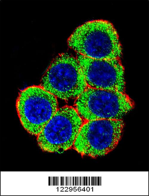
Confocal immunofluorescent analysis of XRCC6 Antibody (Center)(Cat#AP5089c) with 293 cell followed by Alexa Fluor 488-conjugated goat anti-rabbit lgG (green). Actin filaments have been labeled with Alexa Fluor 555 phalloidin (red).DAPI was used to stain the cell nuclear (blue).| Product Name | XRCC6 Antibody (Center) |
|---|---|
| Antibody Type | Primary Antibodies |
| Antigen Alias | XRCC6; G22P1; X-ray repair cross-complementing protein 6; 5'-deoxyribose-5-phosphate lyase Ku70; 70 kDa subunit of Ku antigen; ATP-dependent DNA helicase 2 subunit 1; ATP-dependent DNA helicase II 70 kDa subunit; CTC box-binding factor 75 kDa subunit; DNA |
| Clonality | Polyclonal |
|---|---|
| Isotype | Ig |
| Host Species | Rabbit |
| Tested Applications | WBIHCFCIF |
| WB:1:100~500 IHC |
|
| Species Reactivity | Human |
| Concentration | 1mg/ml |
| Gene Synonyms | G22P1 |
|---|---|
| Alternative Names | XRCC6 G22P1 X-ray repair cross-complementing protein 6 5'-deoxyribose-5-phosphate lyase Ku70 70 kDa subunit of Ku antigen ATP-dependent DNA helicase 2 subunit 1 ATP-dependent DNA helicase II 70 kDa subunit CTC box-binding factor 75 kDa subunit DNA |
| Molecular Weight(MW) | 69843 Da |
| Function | Single stranded DNA-dependent ATP-dependent helicase. Has a role in chromosome translocation. The DNA helicase II complex binds preferentially to fork-like ends of double-stranded DNA in a cell cycle-dependent manner. It works in the 3'-5' direction. Binding to DNA may be mediated by XRCC6. Involved in DNA non-homologous end joining (NHEJ) required for double-strand break repair and V(D)J recombination. The XRCC5/6 dimer acts as regulatory subunit of the DNA-dependent protein kinase complex DNA-PK by increasing the affinity of the catalytic subunit PRKDC to DNA by 100-fold. The XRCC5/6 dimer is probably involved in stabilizing broken DNA ends and bringing them together. The assembly of the DNA-PK complex to DNA ends is required for the NHEJ ligation step. Required for osteocalcin gene expression Probably also acts as a 5'-deoxyribose-5-phosphate lyase (5'-dRP lyase), by catalyzing the beta-elimination of the 5' deoxyribose- 5-phosphate at an abasic site near double-strand breaks. 5'-dRP lyase activity allows to 'clean' the termini of abasic sites, a class of nucleotide damage commonly associated with strand breaks, before such broken ends can be joined. The XRCC5/6 dimer together with APEX1 acts as a negative regulator of transcription |
| Tissue Specificity | This XRCC6 antibody is generated from rabbits immunized with a KLH conjugated synthetic peptide between 235-262 amino acids from the Central region of human XRCC6. |
| Cellular Localization | Nucleus. Chromosome. |
| Entrez Gene | 2547 |
|---|---|
| Nucleotide Accession | NP_001460.1 |
Application
XRCC6 Antibody (Center) western blot analysis in Hela cell line lysates (35ug/lane).This demonstrates the XRCC6 antibody detected the XRCC6 protein (arrow).
Application
XRCC6 antibody (Center) immunohistochemistry analysis in formalin fixed and paraffin embedded human brain tissue followed by peroxidase conjugation of the secondary antibody and DAB staining. This data demonstrates the use of the XRCC6 antibody (Center) for immunohistochemistry. Clinical relevance has not been evaluated.
Application
Western blot analysis of XRCC6 (arrow) using rabbit polyclonal XRCC6 Antibody (Center) . 293 cell lysates (2 ug/lane) either nontransfected (Lane 1) or transiently transfected (Lane 2) with the XRCC6 gene.
Application
XRCC6 Antibody (Center) flow cytometric analysis of Hela cells (right histogram) compared to a negative control cell (left histogram).FITC-conjugated goat-anti-rabbit secondary antibodies were used for the analysis.
Application
Confocal immunofluorescent analysis of XRCC6 Antibody (Center)(Cat#AP5089c) with 293 cell followed by Alexa Fluor 488-conjugated goat anti-rabbit lgG (green). Actin filaments have been labeled with Alexa Fluor 555 phalloidin (red).DAPI was used to stain the cell nuclear (blue).| Application Notes | WB:1:100~500 IHC |
|---|
| Form | Liquid |
|---|---|
| Storage Instructions | For short-term storage, store at 4° C. For long-term storage, aliquot and store at -20ºC or below. Avoid multiple freeze-thaw cycles. |
| Storage Buffer | Purified polyclonal antibody supplied in PBS with 0.09% (W/V) sodium azide. This antibody is purified through a protein A column, followed by peptide affinity purification. |


