Application
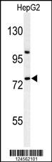
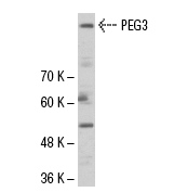
Western blot analysis of ZNF202 Antibody (Center) in HepG2 cell line lysates (35ug/lane). ZNF202 (arrow) was detected using the purified Pab.Application
ZNF202 Antibody (Center) IHC analysis in formalin fixed and paraffin embedded breast carcinoma followed by peroxidase conjugation of the secondary antibody and DAB staining. This data demonstrates the use of the ZNF202 Antibody (Center) for immunohistochemistry. Clinical relevance has not been evaluated.Application
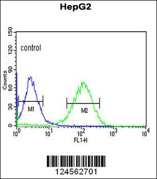
ZNF202 Antibody (Center) flow cytometric analysis of HepG2 cells (right histogram) compared to a negative control cell (left histogram).FITC-conjugated goat-anti-rabbit secondary antibodies were used for the analysis.Application
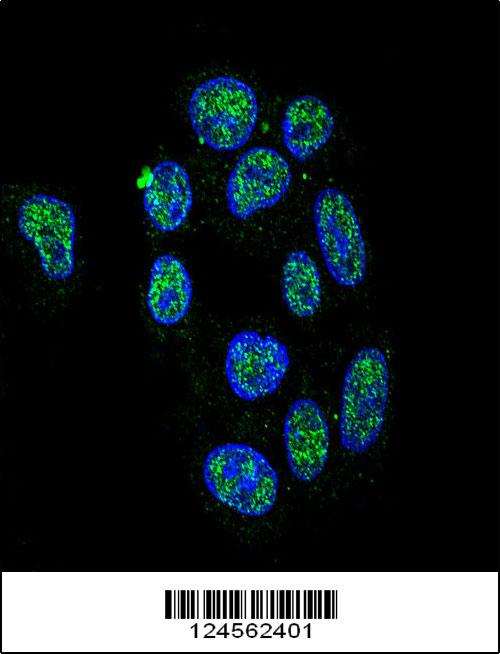
Confocal immunofluorescent analysis of ZNF202 Antibody (Center) (Cat#AP9774c) with HepG2 cell followed by Alexa Fluor 488-conjugated goat anti-rabbit lgG (green). DAPI was used to stain the cell nuclear (blue).Application
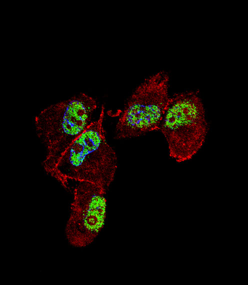
Fluorescent confocal image of MDA-MB231 cell stained with ZNF202 Antibody (Center)(Cat#AP9774c). MDA-MB231 cells were fixed with 4% PFA (20 min), permeabilized with Triton X-100 (0.1%, 10 min), then incubated with ZNF202 primary antibody (1:25, 1 h at 37℃). For secondary antibody, Alexa Fluor® 488 conjugated donkey anti-rabbit antibody (green) was used (1:400, 50 min at 37℃).Cytoplasmic actin was counterstained with Alexa Fluor® 555 (red) conjugated Phalloidin (7units/ml, 1 h at 37℃). Nuclei were counterstained with DAPI (blue) (10 µg/ml, 10 min).ZNF202 immunoreactivity is localized to nucleus significantly.| Product Name | ZNF202 Antibody (Center) |
|---|---|
| Antibody Type | Primary Antibodies |
| Antigen Alias | ZNF202; ZKSCAN10; Zinc finger protein 202; Zinc finger protein with KRAB and SCAN domains 10 |
| Clonality | Polyclonal |
|---|---|
| Isotype | Ig |
| Host Species | Rabbit |
| Tested Applications | WBIHCFCIF |
| WB:1:100~500 IHC |
|
| Species Reactivity | Human |
| Concentration | 1mg/ml |
| Gene Synonyms | ZKSCAN10 |
|---|---|
| Alternative Names | ZNF202 ZKSCAN10 Zinc finger protein 202 Zinc finger protein with KRAB and SCAN domains 10 |
| Molecular Weight(MW) | 74720 Da |
| Function | Transcriptional repressor that binds to elements found predominantly in genes that participate in lipid metabolism. Among its targets are structural components of lipoprotein particles (apolipoproteins AIV, CIII, and E), enzymes involved in lipid processing (lipoprotein lipase, lecithin cholesteryl ester transferase), transporters involved in lipid homeostasis (ABCA1, ABCG1), and several genes involved in processes related to energy metabolism and vascular disease |
| Tissue Specificity | Highly expressed in testis. Also expressed in breast carcinoma cell lines |
| Cellular Localization | Nucleus. |
| Entrez Gene | 7753 |
|---|
Application
Western blot analysis of ZNF202 Antibody (Center) in HepG2 cell line lysates (35ug/lane). ZNF202 (arrow) was detected using the purified Pab.
Application
ZNF202 Antibody (Center) IHC analysis in formalin fixed and paraffin embedded breast carcinoma followed by peroxidase conjugation of the secondary antibody and DAB staining. This data demonstrates the use of the ZNF202 Antibody (Center) for immunohistochemistry. Clinical relevance has not been evaluated.
Application
ZNF202 Antibody (Center) flow cytometric analysis of HepG2 cells (right histogram) compared to a negative control cell (left histogram).FITC-conjugated goat-anti-rabbit secondary antibodies were used for the analysis.
Application
Confocal immunofluorescent analysis of ZNF202 Antibody (Center) (Cat#AP9774c) with HepG2 cell followed by Alexa Fluor 488-conjugated goat anti-rabbit lgG (green). DAPI was used to stain the cell nuclear (blue).
Application
Fluorescent confocal image of MDA-MB231 cell stained with ZNF202 Antibody (Center)(Cat#AP9774c). MDA-MB231 cells were fixed with 4% PFA (20 min), permeabilized with Triton X-100 (0.1%, 10 min), then incubated with ZNF202 primary antibody (1:25, 1 h at 37℃). For secondary antibody, Alexa Fluor® 488 conjugated donkey anti-rabbit antibody (green) was used (1:400, 50 min at 37℃).Cytoplasmic actin was counterstained with Alexa Fluor® 555 (red) conjugated Phalloidin (7units/ml, 1 h at 37℃). Nuclei were counterstained with DAPI (blue) (10 µg/ml, 10 min).ZNF202 immunoreactivity is localized to nucleus significantly.| Application Notes | WB:1:100~500 IHC |
|---|
| Form | Liquid |
|---|---|
| Storage Instructions | For short-term storage, store at 4° C. For long-term storage, aliquot and store at -20ºC or below. Avoid multiple freeze-thaw cycles. |
| Storage Buffer | Purified polyclonal antibody supplied in PBS with 0.09% (W/V) sodium azide. This antibody is purified through a protein A column, followed by peptide affinity purification. |


