Application
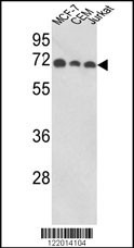
Western blot analysis of ABI1 Antibody (N-term) in MCF-7, CEM, Jurkat cell line lysates (35ug/lane). ABI1 (arrow) was detected using the purified Pab.Application
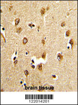
Formalin-fixed and paraffin-embedded human brain tissue reacted with ABI1 Antibody (N-term), which was peroxidase-conjugated to the secondary antibody, followed by DAB staining. This data demonstrates the use of this antibody for immunohistochemistry; clinical relevance has not been evaluated.Application
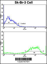
Flow cytometric analysis of SK-Br-3 cells using ABI1 Antibody (N-term)(bottom histogram) compared to a negative control cell (top histogram). FITC-conjugated goat-anti-rabbit secondary antibodies were used for the analysis.Application
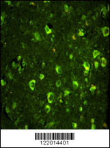
Immunofluorescence analysis of ABI1 Antibody (N-term) with paraffin-embedded human brain tissue . 0.05 mg/ml primary antibody was followed by FITC-conjugated goat anti-rabbit lgG (whole molecule). FITC emits green fluorescence.| Product Name | ABI1 Antibody (N-term) |
|---|---|
| Antibody Type | Primary Antibodies |
| Antigen Alias | ABI1; SSH3BP1; Abl interactor 1; Abelson interactor 1; Abl-binding protein 4; Eps8 SH3 domain-binding protein; Nap1-binding protein; Spectrin SH3 domain-binding protein 1; e3B1 |
| Clonality | Polyclonal |
|---|---|
| Isotype | Ig |
| Host Species | Rabbit |
| Tested Applications | WBIHCFCIF |
| WB:1:100~500 IHC |
|
| Species Reactivity | Human |
| Concentration | 1mg/ml |
| Gene Synonyms | SSH3BP1 |
|---|---|
| Alternative Names | ABI1 SSH3BP1 Abl interactor 1 Abelson interactor 1 Abl-binding protein 4 Eps8 SH3 domain-binding protein Nap1-binding protein Spectrin SH3 domain-binding protein 1 e3B1 |
| Molecular Weight(MW) | 55081 Da |
| Function | May act in negative regulation of cell growth and transformation by interacting with nonreceptor tyrosine kinases ABL1 and/or ABL2. May play a role in regulation of EGF-induced Erk pathway activation. Involved in cytoskeletal reorganization and EGFR signaling. Together with EPS8 participates in transduction of signals from Ras to Rac. In vitro, a trimeric complex of ABI1, EPS8 and SOS1 exhibits Rac specific guanine nucleotide exchange factor (GEF) activity and ABI1 seems to act as an adapter in the complex. Regulates ABL1-mediated phosphorylation of ENAH. Recruits WASF1 to lamellipodia and there seems to regulate WASF1 protein level (By similarity) |
| Tissue Specificity | Widely expressed, with highest expression in brain |
| Cellular Localization | Cytoplasm (By similarity). Nucleus (By similarity). Cell projection, lamellipodium (By similarity). Cell projection, filopodium (By similarity). Cell projection, growth cone (By similarity). Cell junction, synapse, synaptosome (By similarity). Cytoplasm, cytoskeleton (By similarity) Note=Localized to protruding lamellipodia and filopodia tips. Also localized to neuronal growth cones and synaptosomes (By similarity) |
| Entrez Gene | 10006 |
|---|
Application
Western blot analysis of ABI1 Antibody (N-term) in MCF-7, CEM, Jurkat cell line lysates (35ug/lane). ABI1 (arrow) was detected using the purified Pab.
Application
Formalin-fixed and paraffin-embedded human brain tissue reacted with ABI1 Antibody (N-term), which was peroxidase-conjugated to the secondary antibody, followed by DAB staining. This data demonstrates the use of this antibody for immunohistochemistry; clinical relevance has not been evaluated.
Application
Flow cytometric analysis of SK-Br-3 cells using ABI1 Antibody (N-term)(bottom histogram) compared to a negative control cell (top histogram). FITC-conjugated goat-anti-rabbit secondary antibodies were used for the analysis.
Application
Immunofluorescence analysis of ABI1 Antibody (N-term) with paraffin-embedded human brain tissue . 0.05 mg/ml primary antibody was followed by FITC-conjugated goat anti-rabbit lgG (whole molecule). FITC emits green fluorescence.| Application Notes | WB:1:100~500 IHC |
|---|
| Form | Liquid |
|---|---|
| Storage Instructions | For short-term storage, store at 4° C. For long-term storage, aliquot and store at -20ºC or below. Avoid multiple freeze-thaw cycles. |
| Storage Buffer | Purified polyclonal antibody supplied in PBS with 0.09% (W/V) sodium azide. This antibody is purified through a protein A column, followed by peptide affinity purification. |


