Application
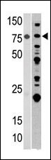
Western blot analysis of anti-ACO2 Pab in mouse heart(left) and 293 (right) tissue lysates (35ug/lane). ACO2(arrow) was detected using the purified Pab (1:60 dilution).Application
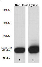
Perfused isolated rat heart whole tissue lysate was lysed with either A) 50 mM Tris-HCl, 150 mM NaCl, 1 mM EDTA, 1% NP-40, 0.1% SDS, 0.5% Na-deoxycholate, 1 mM Na3VO4, 20 mM NaF, 1 mM PMSF, 5 v/v % protease inhibitor cocktail or B) T-PER Tissue Protein Extraction Reagent [# 785101; Pierce], containing 1mM Na3VO4, 20 mM NaF, 5 v/v % protease inhibitor cocktail (Sigma); PVDF membrane was incubated in primary Ab [rabbit polyclonal antibody against ACO2 (Center) (Cat# AP1936c). Solution: 1:1000 diluted in 5% NFM TBS-T 0,05 for overnight (15 hrs) at 4 ?. Data courtesy of Boglarka Laczy M.D., Division of Cardiovascular Disease, Dept. of Medicine, University of Alabama at Birmingham.Application
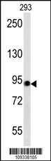
ACO2 western blot analysis in 293 cell line lysates (35ug/lane).This demonstrates the Aconitase antibody detected the Aconitase protein (arrow).Application
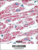
Formalin-fixed and paraffin-embedded human Heart tissue reacted with ACO2 Antibody (Center), which was peroxidase-conjugated to the secondary antibody, followed by AEC staining. This data demonstrates the use of this antibody for immunohistochemistry; clinical relevance has not been evaluated.| Product Name | ACO2 Antibody (Center) |
|---|---|
| Antibody Type | Primary Antibodies |
| Antigen Alias | ACO2; Aconitate hydratase, mitochondrial; Citrate hydro-lyase |
| Clonality | Polyclonal |
|---|---|
| Isotype | Ig |
| Host Species | Rabbit |
| Tested Applications | WBIHC |
| WB:1:100~500 IHC |
|
| Species Reactivity | Human |
| Concentration | 1mg/ml |
| Alternative Names | ACO2 Aconitate hydratase mitochondrial Citrate hydro-lyase |
|---|---|
| Molecular Weight(MW) | 85425 Da |
| Function | Catalyzes the isomerization of citrate to isocitrate via cis-aconitate (By similarity) |
| Tissue Specificity | This ACO2 antibody is generated from rabbits immunized with a KLH conjugated synthetic peptide between 445~475 amino acids from the central region of human Aconitase. |
| Cellular Localization | Mitochondrion (By similarity). |
| Entrez Gene | 50 |
|---|
Application
Western blot analysis of anti-ACO2 Pab in mouse heart(left) and 293 (right) tissue lysates (35ug/lane). ACO2(arrow) was detected using the purified Pab (1:60 dilution).
Application
Perfused isolated rat heart whole tissue lysate was lysed with either A) 50 mM Tris-HCl, 150 mM NaCl, 1 mM EDTA, 1% NP-40, 0.1% SDS, 0.5% Na-deoxycholate, 1 mM Na3VO4, 20 mM NaF, 1 mM PMSF, 5 v/v % protease inhibitor cocktail or B) T-PER Tissue Protein Extraction Reagent [# 785101; Pierce], containing 1mM Na3VO4, 20 mM NaF, 5 v/v % protease inhibitor cocktail (Sigma); PVDF membrane was incubated in primary Ab [rabbit polyclonal antibody against ACO2 (Center) (Cat# AP1936c). Solution: 1:1000 diluted in 5% NFM TBS-T 0,05 for overnight (15 hrs) at 4 ?. Data courtesy of Boglarka Laczy M.D., Division of Cardiovascular Disease, Dept. of Medicine, University of Alabama at Birmingham.
Application
ACO2 western blot analysis in 293 cell line lysates (35ug/lane).This demonstrates the Aconitase antibody detected the Aconitase protein (arrow).
Application
Formalin-fixed and paraffin-embedded human Heart tissue reacted with ACO2 Antibody (Center), which was peroxidase-conjugated to the secondary antibody, followed by AEC staining. This data demonstrates the use of this antibody for immunohistochemistry; clinical relevance has not been evaluated.| Application Notes | WB:1:100~500 IHC |
|---|
| Form | Liquid |
|---|---|
| Storage Instructions | For short-term storage, store at 4° C. For long-term storage, aliquot and store at -20ºC or below. Avoid multiple freeze-thaw cycles. |
| Storage Buffer | Purified polyclonal antibody supplied in PBS with 0.09% (W/V) sodium azide. This antibody is purified through a protein G column, eluted with high and low pH buffers and neutralized immediately, followed by dialysis against PBS. |


