Application
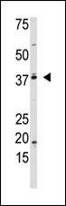
The AP-1 polyclonal antibody is used in Western blot to detect nonsumoylated AP-1 in Jurkat cell lysate.Application
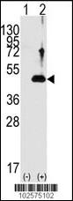
Western blot analysis of AP-1(arrow) using rabbit polyclonal AP-1 Antibody . 293 cell lysates (2 ug/lane) either nontransfected (Lane 1) or transiently transfected with the AP-1 gene (Lane 2) (Origene Technologies).Application
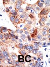
Formalin-fixed and paraffin-embedded human cancer tissue reacted with the primary antibody, which was peroxidase-conjugated to the secondary antibody, followed by DAB staining. This data demonstrates the use of this antibody for immunohistochemistry; clinical relevance has not been evaluated. BC = breast carcinoma; HC = hepatocarcinoma.Application
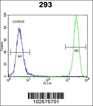
AP-1 Antibody flow cytometric analysis of 293 cells (right histogram) compared to a negative control cell (left histogram).FITC-conjugated goat-anti-rabbit secondary antibodies were used for the analysis.| Product Name | AP-1 Sumoylation Site Antibody |
|---|---|
| Antibody Type | Primary Antibodies |
| Antigen Alias | JUN; Transcription factor AP-1; Activator protein 1; Proto-oncogene c-Jun; V-jun avian sarcoma virus 17 oncogene homolog; p39 |
| Clonality | Polyclonal |
|---|---|
| Isotype | Ig |
| Host Species | Rabbit |
| Tested Applications | WBIHCFC |
| WB:1:100~500 IHC |
|
| Species Reactivity | Human |
| Concentration | 1mg/ml |
| Alternative Names | JUN Transcription factor AP-1 Activator protein 1 Proto-oncogene c-Jun V-jun avian sarcoma virus 17 oncogene homolog p39 |
|---|---|
| Molecular Weight(MW) | 35676 Da |
| Function | Transcription factor that recognizes and binds to the enhancer heptamer motif 5'-TGA[CG]TCA-3'. Promotes activity of NR5A1 when phosphorylated by HIPK3 leading to increased steroidogenic gene expression upon cAMP signaling pathway stimulation |
| Tissue Specificity | This AP-1 Sumoylation Site antibody is generated from rabbits immunized with the KLH conjugated synthetic peptide HPRLQALKEEPQTVPE containing a predicted sumoylation site from the C-terminal region of human AP-1. |
| Cellular Localization | Nucleus. |
| Entrez Gene | 3725 |
|---|
Application
The AP-1 polyclonal antibody is used in Western blot to detect nonsumoylated AP-1 in Jurkat cell lysate.
Application
Western blot analysis of AP-1(arrow) using rabbit polyclonal AP-1 Antibody . 293 cell lysates (2 ug/lane) either nontransfected (Lane 1) or transiently transfected with the AP-1 gene (Lane 2) (Origene Technologies).
Application
Formalin-fixed and paraffin-embedded human cancer tissue reacted with the primary antibody, which was peroxidase-conjugated to the secondary antibody, followed by DAB staining. This data demonstrates the use of this antibody for immunohistochemistry; clinical relevance has not been evaluated. BC = breast carcinoma; HC = hepatocarcinoma.
Application
AP-1 Antibody flow cytometric analysis of 293 cells (right histogram) compared to a negative control cell (left histogram).FITC-conjugated goat-anti-rabbit secondary antibodies were used for the analysis.| Application Notes | WB:1:100~500 IHC |
|---|
| Form | Liquid |
|---|---|
| Storage Instructions | For short-term storage, store at 4° C. For long-term storage, aliquot and store at -20ºC or below. Avoid multiple freeze-thaw cycles. |
| Storage Buffer | Purified polyclonal antibody supplied in PBS with 0.09% (W/V) sodium azide. This antibody is purified through a protein G column, eluted with high and low pH buffers and neutralized immediately, followed by dialysis against PBS. |


