Application
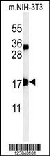
Western blot analysis of APITD1 Antibody (Center) in mouse NIH-3T3 cell line lysates (35ug/lane). APITD1 (arrow) was detected using the purified Pab.Application
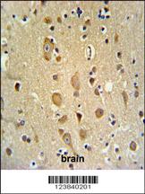
APITD1 Antibody (Center) IHC analysis in formalin fixed and paraffin embedded brain tissue followed by peroxidase conjugation of the secondary antibody and DAB staining. This data demonstrates the use of the APITD1 Antibody (Center) for immunohistochemistry. Clinical relevance has not been evaluated.Application
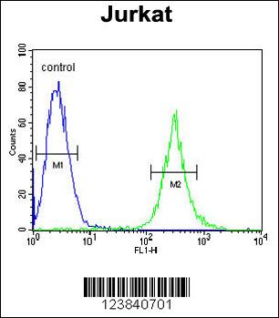
APITD1 Antibody (Center) flow cytometric analysis of Jurkat cells (right histogram) compared to a negative control cell (left histogram).FITC-conjugated goat-anti-rabbit secondary antibodies were used for the analysis.| Product Name | APITD1 Antibody (Center) |
|---|---|
| Antibody Type | Primary Antibodies |
| Antigen Alias | APITD1; CENPS; FAAP16; MHF1; Centromere protein S; Apoptosis-inducing TAF9-like domain-containing protein 1; FANCM-interacting histone fold protein 1; Fanconi anemia-associated polypeptide of 16 kDa |
| Clonality | Polyclonal |
|---|---|
| Isotype | Ig |
| Host Species | Rabbit |
| Tested Applications | WBIHCFC |
| WB:1:100~500 IHC |
|
| Species Reactivity | Human |
| Concentration | 1mg/ml |
| Gene Synonyms | CENPS FAAP16 MHF1 |
|---|---|
| Alternative Names | APITD1 CENPS FAAP16 MHF1 Centromere protein S Apoptosis-inducing TAF9-like domain-containing protein 1 FANCM-interacting histone fold protein 1 Fanconi anemia-associated polypeptide of 16 kDa |
| Molecular Weight(MW) | 15893 Da |
| Function | DNA-binding component of the FA core complex involved in DNA damage repair and genome maintenance. Required for optimal chromatin association of the FA core complex. Required for efficient damage-induced monoubiquitination and focus formation of FANCD2. Stabilizes FAAD24, FANCM and STRA13/CENPX in the FA core complex. Plays a role in DNA interstrand cross-linking (ICL) repair and in recovery of replication forks stalled by topoisomerase I-DNA cleavage intermediates induced by camptothecin. Component of the heterotetrameric CENP-T-W-S-X complex that binds and supercoils DNA, and plays an important role in kinetochore assembly. Component of the APITD1/CENPS complex that is essential for the stable assembly of the outer kinetochore. Plays an important role in mitotic progression and chromosome segregation. Component of the CENPA-CAD (nucleosome distal) complex, a complex recruited to centromeres which is involved in assembly of kinetochore proteins, mitotic progression and chromosome segregation |
| Tissue Specificity | Ubiquitously expressed. |
| Cellular Localization | Nucleus. Chromosome, centromere. Chromosome, centromere, kinetochore. Note=Constitutively localizes to centromeres throughout the cell cycle, and to kinetochores during mitosis. The CENPA-CAD complex is probably recruited on centromeres by the CENPA-NAC complex |
| Entrez Gene | 378708 |
|---|
Application
Western blot analysis of APITD1 Antibody (Center) in mouse NIH-3T3 cell line lysates (35ug/lane). APITD1 (arrow) was detected using the purified Pab.
Application
APITD1 Antibody (Center) IHC analysis in formalin fixed and paraffin embedded brain tissue followed by peroxidase conjugation of the secondary antibody and DAB staining. This data demonstrates the use of the APITD1 Antibody (Center) for immunohistochemistry. Clinical relevance has not been evaluated.
Application
APITD1 Antibody (Center) flow cytometric analysis of Jurkat cells (right histogram) compared to a negative control cell (left histogram).FITC-conjugated goat-anti-rabbit secondary antibodies were used for the analysis.| Application Notes | WB:1:100~500 IHC |
|---|
| Form | Liquid |
|---|---|
| Storage Instructions | For short-term storage, store at 4° C. For long-term storage, aliquot and store at -20ºC or below. Avoid multiple freeze-thaw cycles. |
| Storage Buffer | Purified polyclonal antibody supplied in PBS with 0.09% (W/V) sodium azide. This antibody is purified through a protein A column, followed by peptide affinity purification. |


