Application
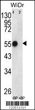
Western blot analysis of ATP5B Antibody (Center) Pab pre-incubated without(lane 1) and with(lane 2) blocking peptide in WiDr cell line lysate. ATP5B (arrow) was detected using the purified Pab (1:60/250 dilution).Application
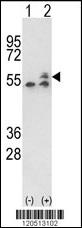
Western blot analysis of ATP5B (arrow) using rabbit polyclonal ATP5B Antibody (Center) . 293 cell lysates (2 ug/lane) either nontransfected (Lane 1) or transiently transfected with the ATP5B gene (Lane 2) .Application
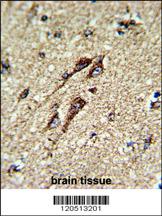
Formalin-fixed and paraffin-embedded human brain tissue reacted with ATP5B Antibody (Center), which was peroxidase-conjugated to the secondary antibody, followed by DAB staining. This data demonstrates the use of this antibody for immunohistochemistry; clinical relevance has not been evaluated.Application
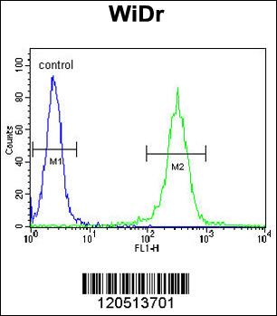
ATP5B Antibody (Center) flow cytometric analysis of WiDr cells (right histogram) compared to a negative control cell (left histogram).FITC-conjugated goat-anti-rabbit secondary antibodies were used for the analysis.| Product Name | ATP5B Antibody (Center) |
|---|---|
| Antibody Type | Primary Antibodies |
| Clonality | Polyclonal |
|---|---|
| Isotype | Ig |
| Host Species | Rabbit |
| Tested Applications | WBIHCFC |
| WB:1:100~500 IHC |
|
| Species Reactivity | Human |
| Concentration | 1mg/ml |
| Gene Synonyms | ATPMB, ATPSB |
|---|---|
| Alternative Names | ATP5B ATPMB ATPSB ATP synthase subunit beta mitochondrial |
| Molecular Weight(MW) | 56560 Da |
| Function | Mitochondrial membrane ATP synthase (F(1)F(0) ATP synthase or Complex V) produces ATP from ADP in the presence of a proton gradient across the membrane which is generated by electron transport complexes of the respiratory chain. F-type ATPases consist of two structural domains, F(1) - containing the extramembraneous catalytic core, and F(0) - containing the membrane proton channel, linked together by a central stalk and a peripheral stalk. During catalysis, ATP synthesis in the catalytic domain of F(1) is coupled via a rotary mechanism of the central stalk subunits to proton translocation. Subunits alpha and beta form the catalytic core in F(1). Rotation of the central stalk against the surrounding alpha(3)beta(3) subunits leads to hydrolysis of ATP in three separate catalytic sites on the beta subunits |
| Tissue Specificity | This ATP5B antibody is generated from rabbits immunized with a KLH conjugated synthetic peptide between 142~171 amino acids from the Center region of human ATP5B. |
| Cellular Localization | Mitochondrion. Mitochondrion inner membrane. Note=Peripheral membrane protein |
| Entrez Gene | 506 |
|---|
Application
Western blot analysis of ATP5B Antibody (Center) Pab pre-incubated without(lane 1) and with(lane 2) blocking peptide in WiDr cell line lysate. ATP5B (arrow) was detected using the purified Pab (1:60/250 dilution).
Application
Western blot analysis of ATP5B (arrow) using rabbit polyclonal ATP5B Antibody (Center) . 293 cell lysates (2 ug/lane) either nontransfected (Lane 1) or transiently transfected with the ATP5B gene (Lane 2) .
Application
Formalin-fixed and paraffin-embedded human brain tissue reacted with ATP5B Antibody (Center), which was peroxidase-conjugated to the secondary antibody, followed by DAB staining. This data demonstrates the use of this antibody for immunohistochemistry; clinical relevance has not been evaluated.
Application
ATP5B Antibody (Center) flow cytometric analysis of WiDr cells (right histogram) compared to a negative control cell (left histogram).FITC-conjugated goat-anti-rabbit secondary antibodies were used for the analysis.| Application Notes | WB:1:100~500 IHC |
|---|
| Form | Liquid |
|---|---|
| Storage Instructions | For short-term storage, store at 4° C. For long-term storage, aliquot and store at -20ºC or below. Avoid multiple freeze-thaw cycles. |
| Storage Buffer | Purified polyclonal antibody supplied in PBS with 0.09% (W/V) sodium azide. This antibody is purified through a protein A column, followed by peptide affinity purification. |


