Application
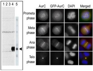
The anti-Aurora C Pab is used in Western blot to detect Aurora C in lysates of 293 cells expressing Flag tag (lane 1), Flag-tagged Aurora A (lane 2), Flag-tagged Aurora B (lane 3), Flag-tagged Aurora C (lane 4), and in A375 cell lysate. In the immunofluorescence experiment, staining of HeLa cells expressing GFP-Aurora C is performed at different cellular mitotic stages with the anti-Aurora C Pab as primary antibody (column A), GFP fluorescence (column B), DAPI nuclear staining (column C), and anti-Aurora C merged to DAPI staining (column D). Data with Flagged Aurora is kindly provided by Drs. K. Sasai and S. Sen from the University of Texas MD Anderson Cancer Center (Houston, TX).Application
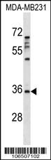
Aurora Antibody (C) western blot analysis in MDA-MB231 cell line lysates (35ug/lane).This demonstrates the Aurora antibody detected the Aurora protein (arrow).| Product Name | Aurora-C Antibody (N-term) |
|---|---|
| Antibody Type | Primary Antibodies |
| Clonality | Polyclonal |
|---|---|
| Isotype | Ig |
| Host Species | Rabbit |
| Tested Applications | WB |
| WB:1:100~500 |
|
| Species Reactivity | Human |
| Concentration | 1mg/ml |
| Gene Synonyms | AIE2, AIK3, AIRK3, ARK3, STK13 |
|---|---|
| Alternative Names | AURKC AIE2 AIK3 AIRK3 ARK3 STK13 Aurora kinase C Aurora 3 Aurora/IPL1-related kinase 3 Aurora/IPL1/Eg2 protein 2 Serine/threonine-protein kinase 13 Serine/threonine-protein kinase aurora-C |
| Molecular Weight(MW) | 35591 Da |
| Function | Serine/threonine-protein kinase component of the chromosomal passenger complex (CPC), a complex that acts as a key regulator of mitosis. The CPC complex has essential functions at the centromere in ensuring correct chromosome alignment and segregation and is required for chromatin-induced microtubule stabilization and spindle assembly. Plays also a role in meiosis and more particularly in spermatogenesis. Has redundant cellular functions with AURKB and can rescue an AURKB knockdown. Like AURKB, AURKC phosphorylates histone H3 at 'Ser-10' and 'Ser-28' Phosphorylates TACC1, another protein involved in cell division, at 'Ser-228' |
| Tissue Specificity | Isoform 1 and isoform 2 are expressed in testis. Elevated expression levels were seen only in a subset of cancer cell lines such as Hep-G2, Huh-7 and HeLa. Expression is maximum at M phase |
| Cellular Localization | Nucleus. Chromosome. Chromosome, centromere. Cytoplasm, cytoskeleton, spindle. Note=Distributes in the condensed chromosomes during prophase to metaphase. After entering anaphase, there is a dissociation from separated chromosomes and a redistribution to midzone microtubules, and finally remains in the midbody during cytokinesis |
| Entrez Gene | 6795 |
|---|
Application
The anti-Aurora C Pab is used in Western blot to detect Aurora C in lysates of 293 cells expressing Flag tag (lane 1), Flag-tagged Aurora A (lane 2), Flag-tagged Aurora B (lane 3), Flag-tagged Aurora C (lane 4), and in A375 cell lysate. In the immunofluorescence experiment, staining of HeLa cells expressing GFP-Aurora C is performed at different cellular mitotic stages with the anti-Aurora C Pab as primary antibody (column A), GFP fluorescence (column B), DAPI nuclear staining (column C), and anti-Aurora C merged to DAPI staining (column D). Data with Flagged Aurora is kindly provided by Drs. K. Sasai and S. Sen from the University of Texas MD Anderson Cancer Center (Houston, TX).
Application
Aurora Antibody (C) western blot analysis in MDA-MB231 cell line lysates (35ug/lane).This demonstrates the Aurora antibody detected the Aurora protein (arrow).| Application Notes | WB:1:100~500 |
|---|
| Form | Liquid |
|---|---|
| Storage Instructions | For short-term storage, store at 4° C. For long-term storage, aliquot and store at -20ºC or below. Avoid multiple freeze-thaw cycles. |
| Storage Buffer | Purified polyclonal antibody supplied in PBS with 0.09% (W/V) sodium azide. This antibody is purified through a protein G column, eluted with high and low pH buffers and neutralized immediately, followed by dialysis against PBS. |


