Application
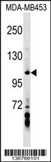
AXIN1 Antibody (Center) western blot analysis in MDA-MB453 cell line lysates (35ug/lane).This demonstrates the AXIN1 antibody detected the AXIN1 protein (arrow).| Product Name | AXIN1 Antibody (Center) |
|---|---|
| Antibody Type | Primary Antibodies |
| Clonality | Polyclonal |
|---|---|
| Isotype | Ig |
| Host Species | Rabbit |
| Tested Applications | WB |
| WB:1:100~500 |
|
| Species Reactivity | Human |
| Concentration | 1mg/ml |
| Gene Synonyms | AXIN |
|---|---|
| Alternative Names | AXIN1 AXIN Axin-1 Axis inhibition protein 1 |
| Molecular Weight(MW) | 95635 Da |
| Function | Component of the beta-catenin destruction complex required for regulating CTNNB1 levels through phosphorylation and ubiquitination, and modulating Wnt-signaling. Controls dorsoventral patterning via two opposing effects; down-regulates CTNNB1 to inhibit the Wnt signaling pathway and ventralize embryos, but also dorsalizes embryos by activating a Wnt- independent JNK signaling pathway. In Wnt signaling, probably facilitates the phosphorylation of CTNNB1 and APC by GSK3B. Likely to function as a tumor suppressor. Facilitates the phosphorylation of TP53 by HIPK2 upon ultraviolet irradiation. Enhances TGF-beta signaling by recruiting the RNF111 E3 ubiquitin ligase and promoting the degradation of inhibitory SMAD7. Also component of the AXIN1-HIPK2-TP53 complex which controls cell growth, apoptosis and development |
| Tissue Specificity | Ubiquitously expressed. |
| Cellular Localization | Cytoplasm. Nucleus. Membrane (By similarity). Cell membrane (By similarity). Note=MACF1 is required for its translocation to cell membrane (By similarity). On UV irradiation, translocates to the nucleus and colocalizes with DAAX |
| Entrez Gene | 8312 |
|---|---|
| Nucleotide Accession | NP_003493.1 NP_851393.1 |
Application
AXIN1 Antibody (Center) western blot analysis in MDA-MB453 cell line lysates (35ug/lane).This demonstrates the AXIN1 antibody detected the AXIN1 protein (arrow).| Application Notes | WB:1:100~500 |
|---|
| Form | Liquid |
|---|---|
| Storage Instructions | For short-term storage, store at 4° C. For long-term storage, aliquot and store at -20ºC or below. Avoid multiple freeze-thaw cycles. |
| Storage Buffer | Purified polyclonal antibody supplied in PBS with 0.09% (W/V) sodium azide. This antibody is purified through a protein A column, followed by peptide affinity purification. |


