Application
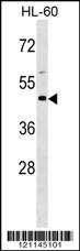
BSG Antibody (Center) western blot analysis in HL-60 cell line lysates (35ug/lane).This demonstrates the BSG antibody detected the BSG protein (arrow).| Product Name | BSG Antibody (Center) |
|---|---|
| Antibody Type | Primary Antibodies |
| Antigen Alias | BSG; Basigin; 5F7; Collagenase stimulatory factor; Extracellular matrix metalloproteinase inducer; Leukocyte activation antigen M6; OK blood group antigen; Tumor cell-derived collagenase stimulatory factor; CD_antigen=CD147; Flags: Precursor |
| Clonality | Polyclonal |
|---|---|
| Isotype | Ig |
| Host Species | Rabbit |
| Tested Applications | WB |
| WB:1:100~500 |
|
| Species Reactivity | Human |
| Concentration | 1mg/ml |
| Alternative Names | BSG Basigin 5F7 Collagenase stimulatory factor Extracellular matrix metalloproteinase inducer Leukocyte activation antigen M6 OK blood group antigen Tumor cell-derived collagenase stimulatory factor CD_antigen=CD147 Flags: Precursor |
|---|---|
| Molecular Weight(MW) | 42200 Da |
| Function | Plays pivotal roles in spermatogenesis, embryo implantation, neural network formation and tumor progression Stimulates adjacent fibroblasts to produce matrix metalloproteinases (MMPS). May target monocarboxylate transporters SLC16A1, SLC16A3 and SLC16A8 to plasma membranes of retinal pigment epithelium and neural retina. Seems to be a receptor for oligomannosidic glycans. In vitro, promotes outgrowth of astrocytic processes (By similarity) |
| Tissue Specificity | Present only in vascular endothelium in non- neoplastic regions of the brain, whereas it is present in tumor cells but not in proliferating blood vessels in malignant gliomas |
| Cellular Localization | Cell membrane; Single-pass type I membrane protein. Melanosome. Note=Colocalizes with SLC16A1 and SLC16A8 (By similarity). Identified by mass spectrometry in melanosome fractions from stage I to stage IV |
| Entrez Gene | 682 |
|---|---|
| Nucleotide Accession | NP_001719.2 NP_940991.1 |
Application
BSG Antibody (Center) western blot analysis in HL-60 cell line lysates (35ug/lane).This demonstrates the BSG antibody detected the BSG protein (arrow).| Application Notes | WB:1:100~500 |
|---|
| Form | Liquid |
|---|---|
| Storage Instructions | For short-term storage, store at 4° C. For long-term storage, aliquot and store at -20ºC or below. Avoid multiple freeze-thaw cycles. |
| Storage Buffer | Purified polyclonal antibody supplied in PBS with 0.09% (W/V) sodium azide. This antibody is purified through a protein A column, followed by peptide affinity purification. |


