Application
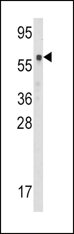
Western blot analysis of CBS Antibody (Center) in mouse kidney tissue lysates (35ug/lane). CBS (arrow) was detected using the purified Pab.Application
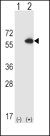
Western blot analysis of CBS (arrow) using rabbit polyclonal CBS Antibody (Center) . 293 cell lysates (2 ug/lane) either nontransfected (Lane 1) or transiently transfected (Lane 2) with the CBS gene.Application
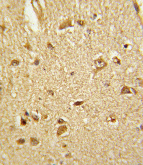
Formalin-fixed and paraffin-embedded human brain tissue reacted with CBS Antibody (Center), which was peroxidase-conjugated to the secondary antibody, followed by DAB staining. This data demonstrates the use of this antibody for immunohistochemistry; clinical relevance has not been evaluated.Application
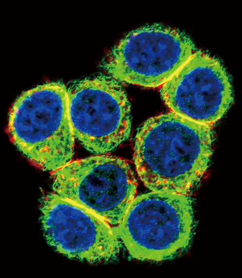
Confocal immunofluorescent analysis of CBS Antibody (Center)(Cat#AP6959c) with 293 cell followed by Alexa Fluor 488-conjugated goat anti-rabbit lgG (green). Actin filaments have been labeled with Alexa Fluor 555 phalloidin (red).DAPI was used to stain the cell nuclear (blue).Application
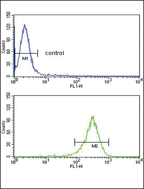
CBS Antibody (Center) flow cytometric analysis of 293 cells (bottom histogram) compared to a negative control cell (top histogram).FITC-conjugated goat-anti-rabbit secondary antibodies were used for the analysis.| Product Name | CBS Antibody (Center) |
|---|---|
| Antibody Type | Primary Antibodies |
| Antigen Alias | CBS; Cystathionine beta-synthase; Beta-thionase; Serine sulfhydrase |
| Clonality | Polyclonal |
|---|---|
| Isotype | Ig |
| Host Species | Rabbit |
| Tested Applications | WBIHCIFFC |
| WB:1:100~500 IHC |
|
| Species Reactivity | Human |
| Concentration | 1mg/ml |
| Alternative Names | CBS Cystathionine beta-synthase Beta-thionase Serine sulfhydrase |
|---|---|
| Molecular Weight(MW) | 60587 Da |
| Function | Only known pyridoxal phosphate-dependent enzyme that contains heme. Important regulator of hydrogen sulfide, especially in the brain, utilizing cysteine instead of serine to catalyze the formation of hydrogen sulfide. Hydrogen sulfide is a gastratransmitter with signaling and cytoprotective effects such as acting as a neuromodulator in the brain to protect neurons against hypoxic injury (By similarity) |
| Tissue Specificity | In the adult strongly expressed in liver and pancreas, some expression in heart and brain, weak expression in lung and kidney. In the fetus, expressed in brain, liver and kidney |
| Cellular Localization | Cytoplasm. Nucleus. |
| Entrez Gene | 875 |
|---|
Application
Western blot analysis of CBS Antibody (Center) in mouse kidney tissue lysates (35ug/lane). CBS (arrow) was detected using the purified Pab.
Application
Western blot analysis of CBS (arrow) using rabbit polyclonal CBS Antibody (Center) . 293 cell lysates (2 ug/lane) either nontransfected (Lane 1) or transiently transfected (Lane 2) with the CBS gene.
Application
Formalin-fixed and paraffin-embedded human brain tissue reacted with CBS Antibody (Center), which was peroxidase-conjugated to the secondary antibody, followed by DAB staining. This data demonstrates the use of this antibody for immunohistochemistry; clinical relevance has not been evaluated.
Application
Confocal immunofluorescent analysis of CBS Antibody (Center)(Cat#AP6959c) with 293 cell followed by Alexa Fluor 488-conjugated goat anti-rabbit lgG (green). Actin filaments have been labeled with Alexa Fluor 555 phalloidin (red).DAPI was used to stain the cell nuclear (blue).
Application
CBS Antibody (Center) flow cytometric analysis of 293 cells (bottom histogram) compared to a negative control cell (top histogram).FITC-conjugated goat-anti-rabbit secondary antibodies were used for the analysis.| Application Notes | WB:1:100~500 IHC |
|---|
| Form | Liquid |
|---|---|
| Storage Instructions | For short-term storage, store at 4° C. For long-term storage, aliquot and store at -20ºC or below. Avoid multiple freeze-thaw cycles. |
| Storage Buffer | Purified polyclonal antibody supplied in PBS with 0.09% (W/V) sodium azide. This antibody is purified through a protein A column, followed by peptide affinity purification. |


