Application
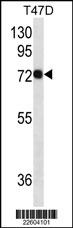
CD138 Antibody (Cat. #AM2157a) western blot analysis in T47D cell line lysates (35ug/lane).This demonstrates the CD138 antibody detected the CD138 protein (arrow).Application
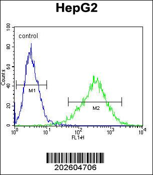
CD138 Antibody flow cytometric analysis of HepG2 cells (right histogram) compared to a negative control cell (left histogram).Alexa Fluor 488-conjugated donkey anti-mouse lgG secondary antibodies were used for the analysisApplication
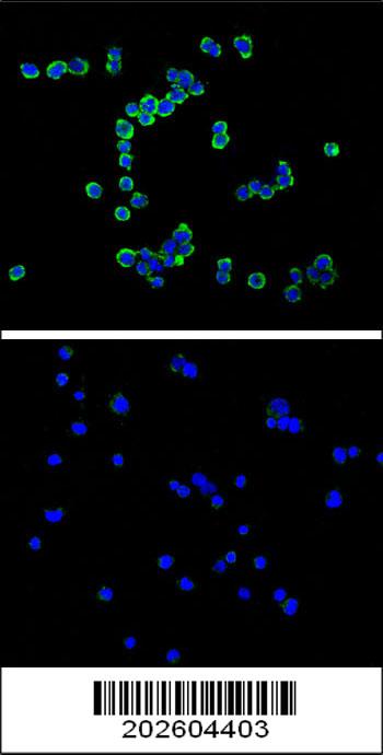
Confocal immunofluorescent analysis of CD138 antibody (Cat#AM2157a) with RPMI8266 cell (above) compared with Jurkat as negative cell line (below).followed by DyLight 488-conjugated goat anti-mouse lgG (H+L) Secondary Antibody (green).DAPI was used to stain the cell nucleus (blue).| Product Name | CD138 Antibody |
|---|---|
| Antibody Type | Primary Antibodies |
| Clonality | Monoclonal |
|---|---|
| Isotype | IgG1 |
| Clone Number | 480CT5.4.3 |
| Host Species | Mouse |
| Tested Applications | WBFCIF |
| WB:1:100~500 FC |
|
| Species Reactivity | Human |
| Concentration | 1mg/ml |
| Gene Synonyms | SDC |
|---|---|
| Alternative Names | SDC1 SDC Syndecan-1 CD_antigen=CD138 Flags: Precursor |
| Molecular Weight(MW) | 32462 Da |
| Function | Cell surface proteoglycan that bears both heparan sulfate and chondroitin sulfate and that links the cytoskeleton to the interstitial matrix |
| Tissue Specificity | This CD138 Monoclonal antibody is generated from mouses immunized with a KLH conjugated synthetic peptide selected from human CD138. |
| Cellular Localization | Membrane; Single-pass type I membrane protein. Secreted. Note=Shedding of the ectodomain produces a soluble form |
| Entrez Gene | 6382 |
|---|
Application
CD138 Antibody (Cat. #AM2157a) western blot analysis in T47D cell line lysates (35ug/lane).This demonstrates the CD138 antibody detected the CD138 protein (arrow).
Application
CD138 Antibody flow cytometric analysis of HepG2 cells (right histogram) compared to a negative control cell (left histogram).Alexa Fluor 488-conjugated donkey anti-mouse lgG secondary antibodies were used for the analysis
Application
Confocal immunofluorescent analysis of CD138 antibody (Cat#AM2157a) with RPMI8266 cell (above) compared with Jurkat as negative cell line (below).followed by DyLight 488-conjugated goat anti-mouse lgG (H+L) Secondary Antibody (green).DAPI was used to stain the cell nucleus (blue).| Application Notes | WB:1:100~500 FC |
|---|
| Form | Liquid |
|---|---|
| Storage Instructions | For short-term storage, store at 4° C. For long-term storage, aliquot and store at -20ºC or below. Avoid multiple freeze-thaw cycles. |
| Storage Buffer | Purified monoclonal antibody supplied in PBS with 0.09% (W/V) sodium azide. This antibody is purified through a protein G column, eluted with high and low pH buffers and neutralized immediately, followed by dialysis against PBS. |


