Application
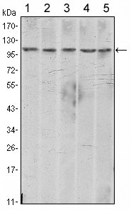
Figure 1: Western blot analysis using CDH2 mouse mAb against A431 (1), NIH/3T3 (2), Hela (3), C6 (4) and LNCap(5) cell lysate.Application
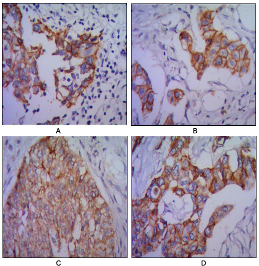
Figure 2: Immunohistochemical analysis of paraffin-embedded human lung cancer (A), colon cancer (B), ovarian cancer??) and mammary cancer(D), using CDH2 mouse mAb with DAB staining.Application
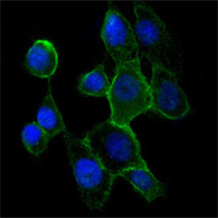
Figure 3:Immunofluorescence analysis of A431 cells using CDH2 mouse mAb (green). Blue: DRAQ5 fluorescent DNA dye.| Product Name | CDH2 Antibody |
|---|---|
| Antibody Type | Primary Antibodies |
| Antigen Alias | CDHN; NCAD; CD325; CDw325; CDH2 |
| Clonality | Monoclonal |
|---|---|
| Isotype | IgG1 |
| Clone Number | 5D5 |
| Host Species | Mouse |
| Tested Applications | WBIHCIF |
| WB:1:500 - 2000. IHC |
|
| Species Reactivity | Human |
| Concentration | 1mg/ml |
| Gene Synonyms | CDHN NCAD |
|---|---|
| Alternative Names | CDHN NCAD CD325 CDw325 CDH2 |
| Molecular Weight(MW) | 99809 Da |
| Function | Cadherins are calcium-dependent cell adhesion proteins. They preferentially interact with themselves in a homophilic manner in connecting cells; cadherins may thus contribute to the sorting of heterogeneous cell types. CDH2 may be involved in neuronal recognition mechanism. In hippocampal neurons, may regulate dendritic spine density (By similarity) |
| Tissue Specificity | Purified recombinant fragment of human CDH2 expressed in E. Coli. |
| Cellular Localization | Cell membrane; Single-pass type I membrane protein |
| Entrez Gene | 1000 |
|---|---|
| Nucleotide Accession | NP_001783.2 |
Application
Figure 1: Western blot analysis using CDH2 mouse mAb against A431 (1), NIH/3T3 (2), Hela (3), C6 (4) and LNCap(5) cell lysate.
Application
Figure 2: Immunohistochemical analysis of paraffin-embedded human lung cancer (A), colon cancer (B), ovarian cancer??) and mammary cancer(D), using CDH2 mouse mAb with DAB staining.
Application
Figure 3:Immunofluorescence analysis of A431 cells using CDH2 mouse mAb (green). Blue: DRAQ5 fluorescent DNA dye.| Application Notes | WB:1:500 - 2000. IHC |
|---|
| Form | Liquid |
|---|---|
| Storage Instructions | For short-term storage, store at 4° C. For long-term storage, aliquot and store at -20ºC or below. Avoid multiple freeze-thaw cycles. |
| Storage Buffer | Purified recombinant fragment of human CDH2 expressed in E. Coli. |


