Application
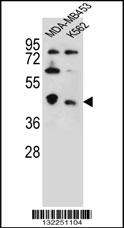
CTBP1 Antibody (C-term) western blot analysis in MDA-MB453,K562 cell line lysates (35ug/lane).This demonstrates the CTBP1 antibody detected the CTBP1 protein (arrow).Application
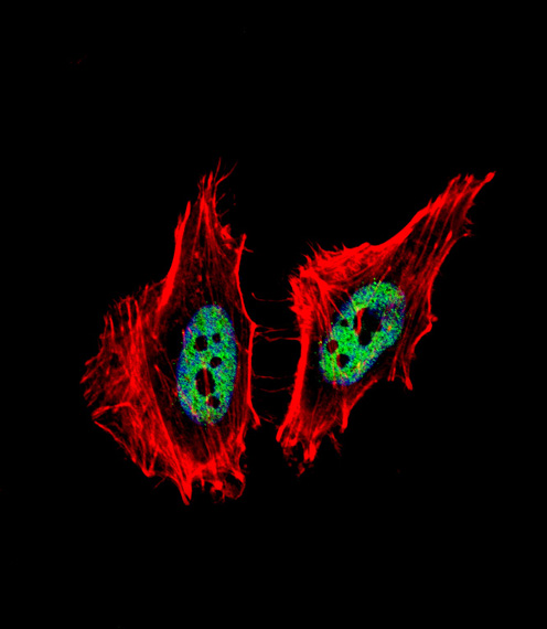
Fluorescent confocal image of Hela cell stained with CTBP1 Antibody (C-term)(Cat#AP16004b). Hela cells were fixed with 4% PFA (20 min), permeabilized with Triton X-100 (0.1%, 10 min), then incubated with CTBP1 primary antibody (1:25, 1 h at 37℃). For secondary antibody, Alexa Fluor® 488 conjugated donkey anti-rabbit antibody (green) was used (1:400, 50 min at 37℃).Cytoplasmic actin was counterstained with Alexa Fluor® 555 (red) conjugated Phalloidin (7units/ml, 1 h at 37℃). Nuclei were counterstained with DAPI (blue) (10 µg/ml, 10 min).CTBP1 immunoreactivity is localized to nucleus significantly.Application
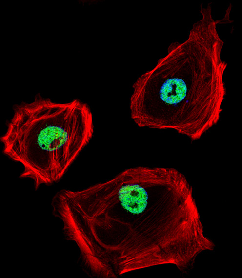
Fluorescent confocal image of SK-BR-3 cell stained with CTBP1 Antibody (C-term)(Cat#AP16004b). SK-BR-3 cells were fixed with 4% PFA (20 min), permeabilized with Triton X-100 (0.1%, 10 min), then incubated with CTBP1 primary antibody (1:25, 1 h at 37℃). For secondary antibody, Alexa Fluor® 488 conjugated donkey anti-rabbit antibody (green) was used (1:400, 50 min at 37℃).Cytoplasmic actin was counterstained with Alexa Fluor® 555 (red) conjugated Phalloidin (7units/ml, 1 h at 37℃). Nuclei were counterstained with DAPI (blue) (10 µg/ml, 10 min).CTBP1 immunoreactivity is localized to nucleus significantly.| Product Name | CTBP1 Antibody (C-term) |
|---|---|
| Antibody Type | Primary Antibodies |
| Antigen Alias | CTBP1; CTBP; C-terminal-binding protein 1 |
| Clonality | Polyclonal |
|---|---|
| Isotype | Ig |
| Host Species | Rabbit |
| Tested Applications | WBIF |
| WB:1:100~500 IF |
|
| Species Reactivity | Human |
| Concentration | 1mg/ml |
| Gene Synonyms | CTBP |
|---|---|
| Alternative Names | CTBP1 CTBP C-terminal-binding protein 1 |
| Molecular Weight(MW) | 47535 Da |
| Function | Involved in controlling the equilibrium between tubular and stacked structures in the Golgi complex. Functions in brown adipose tissue (BAT) differentiation. Corepressor targeting diverse transcription regulators such as GLIS2. Has dehydrogenase activity |
| Tissue Specificity | This CTBP1 antibody is generated from rabbits immunized with a KLH conjugated synthetic peptide between 412-440 amino acids from the C-terminal region of human CTBP1. |
| Cellular Localization | Cytoplasm. Nucleus. |
| Entrez Gene | 1487 |
|---|---|
| Nucleotide Accession | NP_001319.1 NP_001012632.1 |
Application
CTBP1 Antibody (C-term) western blot analysis in MDA-MB453,K562 cell line lysates (35ug/lane).This demonstrates the CTBP1 antibody detected the CTBP1 protein (arrow).
Application
Fluorescent confocal image of Hela cell stained with CTBP1 Antibody (C-term)(Cat#AP16004b). Hela cells were fixed with 4% PFA (20 min), permeabilized with Triton X-100 (0.1%, 10 min), then incubated with CTBP1 primary antibody (1:25, 1 h at 37℃). For secondary antibody, Alexa Fluor® 488 conjugated donkey anti-rabbit antibody (green) was used (1:400, 50 min at 37℃).Cytoplasmic actin was counterstained with Alexa Fluor® 555 (red) conjugated Phalloidin (7units/ml, 1 h at 37℃). Nuclei were counterstained with DAPI (blue) (10 µg/ml, 10 min).CTBP1 immunoreactivity is localized to nucleus significantly.
Application
Fluorescent confocal image of SK-BR-3 cell stained with CTBP1 Antibody (C-term)(Cat#AP16004b). SK-BR-3 cells were fixed with 4% PFA (20 min), permeabilized with Triton X-100 (0.1%, 10 min), then incubated with CTBP1 primary antibody (1:25, 1 h at 37℃). For secondary antibody, Alexa Fluor® 488 conjugated donkey anti-rabbit antibody (green) was used (1:400, 50 min at 37℃).Cytoplasmic actin was counterstained with Alexa Fluor® 555 (red) conjugated Phalloidin (7units/ml, 1 h at 37℃). Nuclei were counterstained with DAPI (blue) (10 µg/ml, 10 min).CTBP1 immunoreactivity is localized to nucleus significantly.| Application Notes | WB:1:100~500 IF |
|---|
| Form | Liquid |
|---|---|
| Storage Instructions | For short-term storage, store at 4° C. For long-term storage, aliquot and store at -20ºC or below. Avoid multiple freeze-thaw cycles. |
| Storage Buffer | Purified polyclonal antibody supplied in PBS with 0.09% (W/V) sodium azide. This antibody is purified through a protein A column, followed by peptide affinity purification. |


