Application
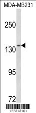
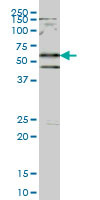
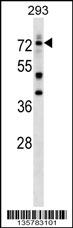
Western blot analysis of CTR9 Antibody (Center) in MDA-MB231 cell line lysates (35ug/lane). CTR9 (arrow) was detected using the purified Pab.Application
(LEFT)Formalin-fixed and paraffin-embedded human brain tissue reacted with CTR9 Antibody (Center), which was peroxidase-conjugated to the secondary antibody, followed by DAB staining. This data demonstrates the use of this antibody for immunohistochemistry; clinical relevance has not been evaluated.(RIGHT)Immunofluorescence analysis of CTR9 Antibody (Center) with paraffin-embedded human brain tissue . 0.05 mg/ml primary antibody was followed by FITC-conjugated goat anti-rabbit lgG (whole molecule). FITC emits green fluorescence.Application
CTR9 Antibody (Center) flow cytometric analysis of k562 cells (bottom histogram) compared to a negative control cell (top histogram).FITC-conjugated goat-anti-rabbit secondary antibodies were used for the analysis.| Product Name | CTR9 Antibody (Center) |
|---|---|
| Antibody Type | Primary Antibodies |
| Antigen Alias | CTR9; KIAA0155; SH2BP1; RNA polymerase-associated protein CTR9 homolog; SH2 domain-binding protein 1 |
| Clonality | Polyclonal |
|---|---|
| Isotype | Ig |
| Host Species | Rabbit |
| Tested Applications | WBIHCFC |
| WB:1:100~500 IHC |
|
| Species Reactivity | Human |
| Concentration | 1mg/ml |
| Gene Synonyms | KIAA0155 SH2BP1 |
|---|---|
| Alternative Names | CTR9 KIAA0155 SH2BP1 RNA polymerase-associated protein CTR9 homolog SH2 domain-binding protein 1 |
| Molecular Weight(MW) | 133502 Da |
| Function | Component of the PAF1 complex (PAF1C) which has multiple functions during transcription by RNA polymerase II and is implicated in regulation of development and maintenance of embryonic stem cell pluripotency. PAF1C associates with RNA polymerase II through interaction with POLR2A CTD non- phosphorylated and 'Ser-2'- and 'Ser-5'-phosphorylated forms and is involved in transcriptional elongation, acting both indepentently and synergistically with TCEA1 and in cooperation with the DSIF complex and HTATSF1. PAF1C is required for transcription of Hox and Wnt target genes. PAF1C is involved in hematopoiesis and stimulates transcriptional activity of MLL1; it promotes leukemogenesis though association with MLL-rearranged oncoproteins, such as MLL-MLLT3/AF9 and MLL-MLLT1/ENL. PAF1C is involved in histone modifications such as ubiquitination of histone H2B and methylation on histone H3 'Lys-4' (H3K4me3). PAF1C recruits the RNF20/40 E3 ubiquitin-protein ligase complex and the E2 enzyme UBE2A or UBE2B to chromatin which mediate monoubiquitination of 'Lys-120' of histone H2B (H2BK120ub1); UB2A/B-mediated H2B ubiquitination is proposed to be coupled to transcription. PAF1C is involved in mRNA 3' end formation probably through association with cleavage and poly(A) factors. In case of infection by influenza A strain H3N2, PAF1C associates with viral NS1 protein, thereby regulating gene transcription. Required for mono- and trimethylation on histone H3 'Lys-4' (H3K4me3) and dimethylation on histone H3 'Lys-79' (H3K4me3). Required for Hox gene transcription. Required for the trimethylation of histone H3 'Lys-4' (H3K4me3) on genes involved in stem cell pluripotency; this function is synergistic with CXXC1 indicative for an involvement of the SET1 complex. Involved in transcriptional regulation of IL6-responsive genes and in JAK-STAT pathway; may regulate DNA-association of STAT3 (By similarity) |
| Tissue Specificity | Widely expressed. |
| Cellular Localization | Nucleus speckle (By similarity). Note=Found in speckles (By similarity) |
| Entrez Gene | 9646 |
|---|
Application
Western blot analysis of CTR9 Antibody (Center) in MDA-MB231 cell line lysates (35ug/lane). CTR9 (arrow) was detected using the purified Pab.
Application
(LEFT)Formalin-fixed and paraffin-embedded human brain tissue reacted with CTR9 Antibody (Center), which was peroxidase-conjugated to the secondary antibody, followed by DAB staining. This data demonstrates the use of this antibody for immunohistochemistry; clinical relevance has not been evaluated.(RIGHT)Immunofluorescence analysis of CTR9 Antibody (Center) with paraffin-embedded human brain tissue . 0.05 mg/ml primary antibody was followed by FITC-conjugated goat anti-rabbit lgG (whole molecule). FITC emits green fluorescence.
Application
CTR9 Antibody (Center) flow cytometric analysis of k562 cells (bottom histogram) compared to a negative control cell (top histogram).FITC-conjugated goat-anti-rabbit secondary antibodies were used for the analysis.| Application Notes | WB:1:100~500 IHC |
|---|
| Form | Liquid |
|---|---|
| Storage Instructions | For short-term storage, store at 4° C. For long-term storage, aliquot and store at -20ºC or below. Avoid multiple freeze-thaw cycles. |
| Storage Buffer | Purified polyclonal antibody supplied in PBS with 0.09% (W/V) sodium azide. This antibody is purified through a protein A column, followed by peptide affinity purification. |


