Application
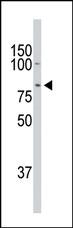
The DCAMKL1 Antibody (C-term ) western blot analysis in mouse heart tissuelysates (35ug/lane).This demonstrates the DCAMKL1 antibody detected the DCAMKL1 protein (arrow).Application
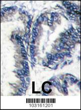
Formalin-fixed and paraffin-embedded human hepatocarcinoma reacted with DCAMKL1 Antibody (C-term), which was peroxidase-conjugated to the secondary antibody, followed by DAB staining. This data demonstrates the use of this antibody for immunohistochemistry; clinical relevance has not been evaluated.Application
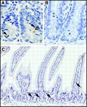
Expression pattern of doublecortin and CaM kinase-like-1 (DCAMKL-1) in normal mouse small intestine. (A): Immunohistochemical staining of normal small intestine for DCAMKL-1. Arrows indicate the cells positive for DCAMKL-1 in the stem cell zone. (B): Preincubation with blocking peptide completely abolished DCAMKL-1 immunoreactivity. (C): Immunohistochemical staining of normal small intestine for DCAMKL-1. Brown indicates the cells positive for DCAMKL-1 (indicated by the arrows).Application
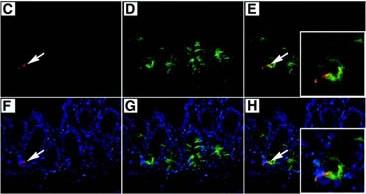
(C): The cell positive for DCAMKL-1, stained red (indicated by the arrow), appears at the base of the crypt. (D): Intestinal section stained for Musashi-1 (green). (E): Colocalization of DCAMKL-1 and Musashi-1 (yellow, indicated by the arrow). The magnified inset image represents the single cell positive for both DCAMKL-1 and Musashi-1. (F): Costaining of DCMAKL-1 (red, indicated by the arrow) with nuclear Hoechst 33342 (blue) staining. (G): Costaining of Musashi-1 (green) with nuclear Hoechst 33342 (blue) staining. (H): Colocalization of DCAMKL-1 and Musashi-1 (yellow, indicated by the arrow), costained with nuclear Hoechst 33342 (blue) staining. The magnified inset image represents the single cell positive for both DCAMKL-1 and Musashi-1 (yellow).Application
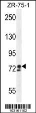
DCAMKL1 Antibody (C-term) western blot analysis in ZR-75-1 cell line lysates (35ug/lane).This demonstrates the DCAMKL1 antibody detected the DCAMKL1 protein (arrow).| Product Name | DCAMKL1 Antibody (C-term) |
|---|---|
| Antibody Type | Primary Antibodies |
| Antigen Alias | DCLK1; DCAMKL1; DCDC3A; KIAA0369; Serine/threonine-protein kinase DCLK1; Doublecortin domain-containing protein 3A; Doublecortin-like and CAM kinase-like 1; Doublecortin-like kinase 1 |
| Clonality | Polyclonal |
|---|---|
| Isotype | Ig |
| Host Species | Rabbit |
| Tested Applications | WBIHCIF |
| WB:1:100~500 IHC |
|
| Species Reactivity | Human |
| Concentration | 1mg/ml |
| Gene Synonyms | DCAMKL1 DCDC3A KIAA0369 |
|---|---|
| Alternative Names | DCLK1 DCAMKL1 DCDC3A KIAA0369 Serine/threonine-protein kinase DCLK1 Doublecortin domain-containing protein 3A Doublecortin-like and CAM kinase-like 1 Doublecortin-like kinase 1 |
| Molecular Weight(MW) | 82224 Da |
| Function | Probable kinase that may be involved in a calcium- signaling pathway controlling neuronal migration in the developing brain. May also participate in functions of the mature nervous system |
| Tissue Specificity | In fetal tissues, highly expressed in brain, detectable in lung and liver, but not in kidney. In adult tissues, expressed ubiquitously in the brain, detectable in the heart, liver, spleen, thymus, prostate, testis, ovary, small intestine and colon. The type A isoforms seem to be expressed predominantly in fetal brain whereas type B isoforms are expressed abundantly in both fetal and adult brain |
| Entrez Gene | 9201 |
|---|
Application
The DCAMKL1 Antibody (C-term ) western blot analysis in mouse heart tissuelysates (35ug/lane).This demonstrates the DCAMKL1 antibody detected the DCAMKL1 protein (arrow).
Application
Formalin-fixed and paraffin-embedded human hepatocarcinoma reacted with DCAMKL1 Antibody (C-term), which was peroxidase-conjugated to the secondary antibody, followed by DAB staining. This data demonstrates the use of this antibody for immunohistochemistry; clinical relevance has not been evaluated.
Application
Expression pattern of doublecortin and CaM kinase-like-1 (DCAMKL-1) in normal mouse small intestine. (A): Immunohistochemical staining of normal small intestine for DCAMKL-1. Arrows indicate the cells positive for DCAMKL-1 in the stem cell zone. (B): Preincubation with blocking peptide completely abolished DCAMKL-1 immunoreactivity. (C): Immunohistochemical staining of normal small intestine for DCAMKL-1. Brown indicates the cells positive for DCAMKL-1 (indicated by the arrows).
Application
(C): The cell positive for DCAMKL-1, stained red (indicated by the arrow), appears at the base of the crypt. (D): Intestinal section stained for Musashi-1 (green). (E): Colocalization of DCAMKL-1 and Musashi-1 (yellow, indicated by the arrow). The magnified inset image represents the single cell positive for both DCAMKL-1 and Musashi-1. (F): Costaining of DCMAKL-1 (red, indicated by the arrow) with nuclear Hoechst 33342 (blue) staining. (G): Costaining of Musashi-1 (green) with nuclear Hoechst 33342 (blue) staining. (H): Colocalization of DCAMKL-1 and Musashi-1 (yellow, indicated by the arrow), costained with nuclear Hoechst 33342 (blue) staining. The magnified inset image represents the single cell positive for both DCAMKL-1 and Musashi-1 (yellow).
Application
DCAMKL1 Antibody (C-term) western blot analysis in ZR-75-1 cell line lysates (35ug/lane).This demonstrates the DCAMKL1 antibody detected the DCAMKL1 protein (arrow).| Application Notes | WB:1:100~500 IHC |
|---|
| Form | Liquid |
|---|---|
| Storage Instructions | For short-term storage, store at 4° C. For long-term storage, aliquot and store at -20ºC or below. Avoid multiple freeze-thaw cycles. |
| Storage Buffer | Purified polyclonal antibody supplied in PBS with 0.09% (W/V) sodium azide. This antibody is purified through a protein G column, eluted with high and low pH buffers and neutralized immediately, followed by dialysis against PBS. |


