Application
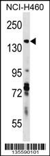
DIAPH3 Antibody (C-term) western blot analysis in NCI-H460 cell line lysates (35ug/lane).This demonstrates the DIAPH3 antibody detected the DIAPH3 protein (arrow).| Product Name | DIAPH3 Antibody (C-term) |
|---|---|
| Antibody Type | Primary Antibodies |
| Antigen Alias | DIAPH3; DIAP3; Protein diaphanous homolog 3; Diaphanous-related formin-3; MDia2 |
| Clonality | Polyclonal |
|---|---|
| Isotype | Ig |
| Host Species | Rabbit |
| Tested Applications | WB |
| WB:1:100~500 |
|
| Species Reactivity | Human |
| Concentration | 1mg/ml |
| Gene Synonyms | DIAP3 |
|---|---|
| Alternative Names | DIAPH3 DIAP3 Protein diaphanous homolog 3 Diaphanous-related formin-3 MDia2 |
| Molecular Weight(MW) | 136926 Da |
| Function | Binds to GTP-bound form of Rho and to profilin. Acts in a Rho-dependent manner to recruit profilin to the membrane, where it promotes actin polymerization. It is required for cytokinesis, stress fiber formation, and transcriptional activation of the serum response factor. DFR proteins couple Rho and Src tyrosine kinase during signaling and the regulation of actin dynamics (By similarity) |
| Tissue Specificity | This DIAPH3 antibody is generated from rabbits immunized with a KLH conjugated synthetic peptide between 1060-1090 amino acids from the C-terminal region of human DIAPH3. |
| Cellular Localization | Cytoplasm, cytosol. Note=During mitosis, co- localizes with the actin-rich cleavage furrow and with the microtubule-rich central spindle during cytokinesis |
| Entrez Gene | 81624 |
|---|---|
| Nucleotide Accession | NP_001035982.1 NP_112194.2 |
Application
DIAPH3 Antibody (C-term) western blot analysis in NCI-H460 cell line lysates (35ug/lane).This demonstrates the DIAPH3 antibody detected the DIAPH3 protein (arrow).| Application Notes | WB:1:100~500 |
|---|
| Form | Liquid |
|---|---|
| Storage Instructions | For short-term storage, store at 4° C. For long-term storage, aliquot and store at -20ºC or below. Avoid multiple freeze-thaw cycles. |
| Storage Buffer | Purified polyclonal antibody supplied in PBS with 0.09% (W/V) sodium azide. This antibody is purified through a protein A column, followed by peptide affinity purification. |


