Application
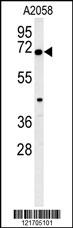
EIF2AK2 Antibody (Center) western blot analysis in A2058 cell line lysates (35ug/lane).This demonstrates the EIF2AK2 antibody detected the EIF2AK2 protein (arrow).Application
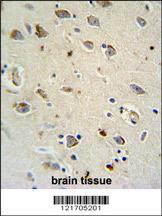
EIF2AK2 Antibody (Center) immunohistochemistry analysis in formalin fixed and paraffin embedded human brain tissue followed by peroxidase conjugation of the secondary antibody and DAB staining. This data demonstrates the use of the EIF2AK2 Antibody (Center) for immunohistochemistry. Clinical relevance has not been evaluated.Application
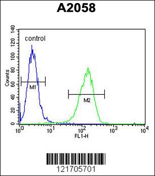
EIF2AK2 Antibody (Center) flow cytometric analysis of A2058 cells (right histogram) compared to a negative control cell (left histogram).FITC-conjugated goat-anti-rabbit secondary antibodies were used for the analysis.Application
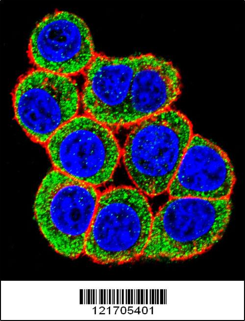
Confocal immunofluorescent analysis of EIF2AK2 Antibody (Center)(Cat#AP5851c) with Hela cell followed by Alexa Fluor 488-conjugated goat anti-rabbit lgG (green). Actin filaments have been labeled with Alexa Fluor 555 phalloidin (red).DAPI was used to stain the cell nuclear (blue).| Product Name | EIF2AK2 Antibody (Center) |
|---|---|
| Antibody Type | Primary Antibodies |
| Clonality | Polyclonal |
|---|---|
| Isotype | Ig |
| Host Species | Rabbit |
| Tested Applications | WBIHCFCIF |
| WB:1:100~500 IHC |
|
| Species Reactivity | Human |
| Concentration | 1mg/ml |
| Gene Synonyms | PKR, PRKR |
|---|---|
| Alternative Names | EIF2AK2 PKR PRKR Interferon-induced double-stranded RNA-activated protein kinase Eukaryotic translation initiation factor 2-alpha kinase 2 Interferon-inducible RNA-dependent protein kinase P1/eIF-2A protein kinase Protein kinase RNA-activated Tyr |
| Molecular Weight(MW) | 62094 Da |
| Function | Following activation by double-stranded RNA in the presence of ATP, the kinase becomes autophosphorylated and can catalyze the phosphorylation of the translation initiation factor EIF2S1, which leads to an inhibition of the initiation of protein synthesis. Double-stranded RNA is generated during the course of a viral infection. In addition to serine/threonine-protein kinase activity, also has tyrosine-protein kinase activity: phosphorylates CDK1 upon DNA damage. CDK1 phosphorylation triggers CDK1 polyubiquitination and subsequent proteolysis, thus leading to G2 arrest |
| Tissue Specificity | This EIF2AK2 antibody is generated from rabbits immunized with a KLH conjugated synthetic peptide between 240-270 amino acids from the Central region of human EIF2AK2. |
| Entrez Gene | 5610 |
|---|---|
| Nucleotide Accession | NP_001129124.1 |
Application
EIF2AK2 Antibody (Center) western blot analysis in A2058 cell line lysates (35ug/lane).This demonstrates the EIF2AK2 antibody detected the EIF2AK2 protein (arrow).
Application
EIF2AK2 Antibody (Center) immunohistochemistry analysis in formalin fixed and paraffin embedded human brain tissue followed by peroxidase conjugation of the secondary antibody and DAB staining. This data demonstrates the use of the EIF2AK2 Antibody (Center) for immunohistochemistry. Clinical relevance has not been evaluated.
Application
EIF2AK2 Antibody (Center) flow cytometric analysis of A2058 cells (right histogram) compared to a negative control cell (left histogram).FITC-conjugated goat-anti-rabbit secondary antibodies were used for the analysis.
Application
Confocal immunofluorescent analysis of EIF2AK2 Antibody (Center)(Cat#AP5851c) with Hela cell followed by Alexa Fluor 488-conjugated goat anti-rabbit lgG (green). Actin filaments have been labeled with Alexa Fluor 555 phalloidin (red).DAPI was used to stain the cell nuclear (blue).| Application Notes | WB:1:100~500 IHC |
|---|
| Form | Liquid |
|---|---|
| Storage Instructions | For short-term storage, store at 4° C. For long-term storage, aliquot and store at -20ºC or below. Avoid multiple freeze-thaw cycles. |
| Storage Buffer | Purified polyclonal antibody supplied in PBS with 0.09% (W/V) sodium azide. This antibody is purified through a protein A column, followed by peptide affinity purification. |


