Application
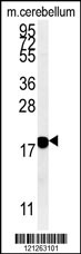
Western blot analysis of anti-EphA4 N-term Pab in HeLa cell lysate. EphA4 (arrow) was detected using purified Pab. Secondary HRP-anti-rabbit was used for signal visualization with chemiluminescence.Application
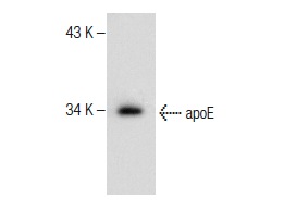
Western blot analysis of EphA4 (arrow) using EphA4 Antibody (N-term) . 293 cell lysates (2 ug/lane) either nontransfected (Lane 1) or transiently transfected with the EphA4 gene (Lane 2) (Origene Technologies).Application
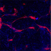
Methanol/Acetone fixed human stem cell is used in IF to detect Eph4A (blue) and endothelial Lectin(red). Data kindly provided by Dr. Weis from Cheresh Lab, UCSD.Application
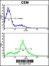
Flow cytometric analysis of CEM cells using EphA4 Antibody (N-term) (bottom histogram) compared to a negative control cell (top histogram). FITC-conjugated goat-anti-rabbit secondary antibodies were used for the analysis.Application
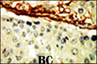
Formalin-fixed and paraffin-embedded human cancer tissue reacted with the primary antibody, which was peroxidase-conjugated to the secondary antibody, followed by DAB staining. This data demonstrates the use of this antibody for immunohistochemistry; clinical relevance has not been evaluated. BC = breast carcinoma; HC = hepatocarcinoma.| Product Name | EphA4 Antibody (N-term) |
|---|---|
| Antibody Type | Primary Antibodies |
| Clonality | Polyclonal |
|---|---|
| Isotype | Ig |
| Host Species | Rabbit |
| Tested Applications | WBIFFCIHC |
| WB:1:100~500 IF |
|
| Species Reactivity | Human |
| Concentration | 1mg/ml |
| Gene Synonyms | HEK8, SEK, TYRO1 |
|---|---|
| Alternative Names | EPHA4 HEK8 SEK TYRO1 Ephrin type-A receptor 4 EPH-like kinase 8 Tyrosine-protein kinase TYRO1 Tyrosine-protein kinase receptor SEK |
| Molecular Weight(MW) | 109860 Da |
| Function | Receptor tyrosine kinase which binds membrane-bound ephrin family ligands residing on adjacent cells, leading to contact-dependent bidirectional signaling into neighboring cells The signaling pathway downstream of the receptor is referred to as forward signaling while the signaling pathway downstream of the ephrin ligand is referred to as reverse signaling. Highly promiscuous, it has the unique property among Eph receptors to bind and to be physiologically activated by both GPI-anchored ephrin-A and transmembrane ephrin-B ligands including EFNA1 and EFNB3. Upon activation by ephrin ligands, modulates cell morphology and integrin-dependent cell adhesion through regulation of the Rac, Rap and Rho GTPases activity. Plays an important role in the development of the nervous system controlling different steps of axonal guidance including the establishment of the corticospinal projections. May also control the segregation of motor and sensory axons during neuromuscular circuit development Beside its role in axonal guidance plays a role in synaptic plasticity. Activated by EFNA1 phosphorylates CDK5 at 'Tyr-15' which in turn phosphorylates NGEF regulating RHOA and dendritic spine morphogenesis. In the nervous system, plays also a role in repair after injury preventing axonal regeneration and in angiogenesis playing a role in central nervous system vascular formation. Additionally, its promiscuity makes it available to participate in a variety of cell-cell signaling regulating for instance the development of the thymic epithelium |
| Tissue Specificity | Ubiquitous. |
| Cellular Localization | Cell membrane; Single-pass type I membrane protein (By similarity). Cell projection, axon (By similarity) Cell projection, dendrite (By similarity). Cell junction, synapse, postsynaptic cell membrane, postsynaptic density (By similarity) Early endosome (By similarity). Note=Clustered upon activation and targeted to early endosome (By similarity) |
| Entrez Gene | 2043 |
|---|
Application
Western blot analysis of anti-EphA4 N-term Pab in HeLa cell lysate. EphA4 (arrow) was detected using purified Pab. Secondary HRP-anti-rabbit was used for signal visualization with chemiluminescence.
Application
Western blot analysis of EphA4 (arrow) using EphA4 Antibody (N-term) . 293 cell lysates (2 ug/lane) either nontransfected (Lane 1) or transiently transfected with the EphA4 gene (Lane 2) (Origene Technologies).
Application
Methanol/Acetone fixed human stem cell is used in IF to detect Eph4A (blue) and endothelial Lectin(red). Data kindly provided by Dr. Weis from Cheresh Lab, UCSD.
Application
Flow cytometric analysis of CEM cells using EphA4 Antibody (N-term) (bottom histogram) compared to a negative control cell (top histogram). FITC-conjugated goat-anti-rabbit secondary antibodies were used for the analysis.
Application
Formalin-fixed and paraffin-embedded human cancer tissue reacted with the primary antibody, which was peroxidase-conjugated to the secondary antibody, followed by DAB staining. This data demonstrates the use of this antibody for immunohistochemistry; clinical relevance has not been evaluated. BC = breast carcinoma; HC = hepatocarcinoma.| Application Notes | WB:1:100~500 IF |
|---|
| Form | Liquid |
|---|---|
| Storage Instructions | For short-term storage, store at 4° C. For long-term storage, aliquot and store at -20ºC or below. Avoid multiple freeze-thaw cycles. |
| Storage Buffer | Purified polyclonal antibody supplied in PBS with 0.09% (W/V) sodium azide. This antibody is purified through a protein G column, eluted with high and low pH buffers and neutralized immediately, followed by dialysis against PBS. |


