Application
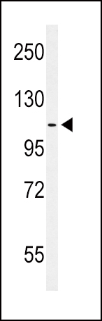
Western blot analysis of hEPHA2-T45 in MCF-7 cell line lysates (35ug/lane). EPHA2 (arrow) was detected using the purified PabApplication
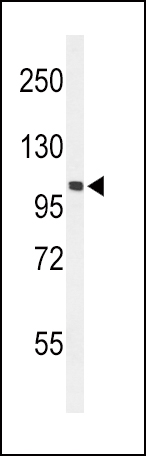
Western blot analysis of hEPHA2-T45 in mouse NIH-3T3 cell line lysates (35ug/lane). EPHA2 (arrow) was detected using the purified Pab.Application
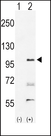
Western blot analysis of EPHA2 (arrow) using rabbit polyclonal hEPHA2-T45 . 293 cell lysates (2 ug/lane) either nontransfected (Lane 1) or transiently transfected with the EPHA2 gene (Lane 2)Application
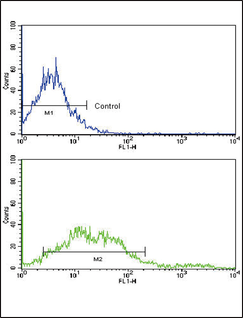
Flow cytometric analysis of NCI-H292 cells using EphA2 Antibody (N-term)(bottom histogram) compared to a negative control cell (top histogram). FITC-conjugated goat-anti-rabbit secondary antibodies were used for the analysis.| Product Name | EphA2 Antibody (N-term) |
|---|---|
| Antibody Type | Primary Antibodies |
| Clonality | Polyclonal |
|---|---|
| Isotype | Ig |
| Host Species | Rabbit |
| Tested Applications | WBFC |
| WB:1:100~500 FC |
|
| Species Reactivity | Human |
| Concentration | 1mg/ml |
| Gene Synonyms | ECK |
|---|---|
| Alternative Names | EPHA2 ECK Ephrin type-A receptor 2 Epithelial cell kinase Tyrosine-protein kinase receptor ECK |
| Molecular Weight(MW) | 108266 Da |
| Function | Receptor tyrosine kinase which binds promiscuously membrane-bound ephrin-A family ligands residing on adjacent cells, leading to contact-dependent bidirectional signaling into neighboring cells. The signaling pathway downstream of the receptor is referred to as forward signaling while the signaling pathway downstream of the ephrin ligand is referred to as reverse signaling. Activated by the ligand ephrin-A1/EFNA1 regulates migration, integrin-mediated adhesion, proliferation and differentiation of cells. Regulates cell adhesion and differentiation through DSG1/desmoglein-1 and inhibition of the ERK1/ERK2 (MAPK3/MAPK1, respectively) signaling pathway. May also participate in UV radiation-induced apoptosis and have a ligand- independent stimulatory effect on chemotactic cell migration During development, may function in distinctive aspects of pattern formation and subsequently in development of several fetal tissues. Involved for instance in angiogenesis, in early hindbrain development and epithelial proliferation and branching morphogenesis during mammary gland development. Engaged by the ligand ephrin-A5/EFNA5 may regulate lens fiber cells shape and interactions and be important for lens transparency development and maintenance. With ephrin-A2/EFNA2 may play a role in bone remodeling through regulation of osteoclastogenesis and osteoblastogenesis |
| Tissue Specificity | Expressed in brain and glioma tissue and glioma cell lines (at protein level). Expressed most highly in tissues that contain a high proportion of epithelial cells, e.g skin, intestine, lung, and ovary |
| Cellular Localization | Cell membrane; Single-pass type I membrane protein. Cell projection, ruffle membrane; Single-pass type I membrane protein. Cell projection, lamellipodium membrane; Single- pass type I membrane protein. Cell junction, focal adhesion Note=Present at regions of cell-cell contacts but also at the leading edge of migrating cells |
| Entrez Gene | 1969 |
|---|
Application
Western blot analysis of hEPHA2-T45 in MCF-7 cell line lysates (35ug/lane). EPHA2 (arrow) was detected using the purified Pab
Application
Western blot analysis of hEPHA2-T45 in mouse NIH-3T3 cell line lysates (35ug/lane). EPHA2 (arrow) was detected using the purified Pab.
Application
Western blot analysis of EPHA2 (arrow) using rabbit polyclonal hEPHA2-T45 . 293 cell lysates (2 ug/lane) either nontransfected (Lane 1) or transiently transfected with the EPHA2 gene (Lane 2)
Application
Flow cytometric analysis of NCI-H292 cells using EphA2 Antibody (N-term)(bottom histogram) compared to a negative control cell (top histogram). FITC-conjugated goat-anti-rabbit secondary antibodies were used for the analysis.| Application Notes | WB:1:100~500 FC |
|---|
| Form | Liquid |
|---|---|
| Storage Instructions | For short-term storage, store at 4° C. For long-term storage, aliquot and store at -20ºC or below. Avoid multiple freeze-thaw cycles. |
| Storage Buffer | Purified polyclonal antibody supplied in PBS with 0.09% (W/V) sodium azide. This antibody is purified through a protein G column, eluted with high and low pH buffers and neutralized immediately, followed by dialysis against PBS. |


