Application
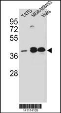
ESR2 Antibody (Center) (Cat. #AX10006) western blot analysis in T47D,MDA-MB453,Hela cell line lysates (35ug/lane).This demonstrates the ESR2 antibody detected the ESR2 protein (arrow).Application
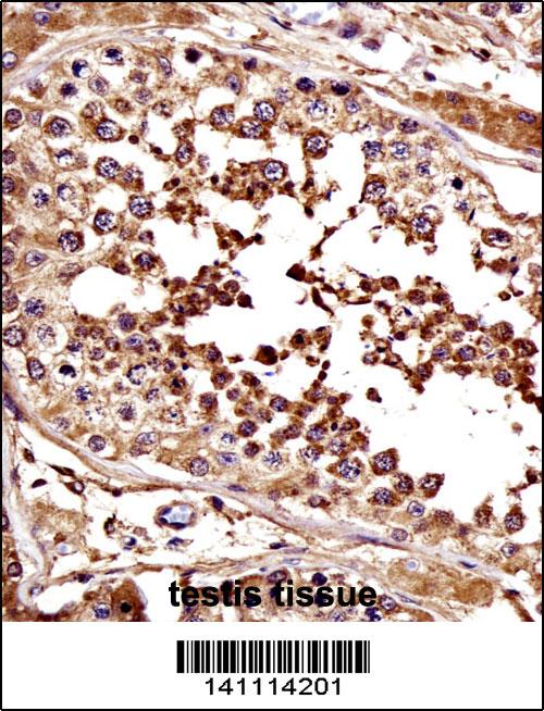
ESR2 Antibody (Center) immunohistochemistry analysis in formalin fixed and paraffin embedded human testis tissue followed by peroxidase conjugation of the secondary antibody and DAB staining.This data demonstrates the use of ESR2 Antibody (Center) for immunohistochemistry. Clinical relevance has not been evaluated.Application
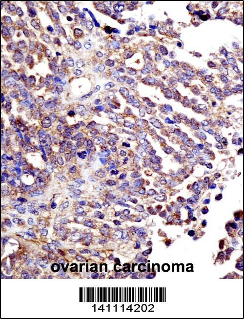
ESR2 Antibody (Center) immunohistochemistry analysis in formalin fixed and paraffin embedded human ovarian carcinoma followed by peroxidase conjugation of the secondary antibody and DAB staining.This data demonstrates the use of ESR2 Antibody (Center) for immunohistochemistry. Clinical relevance has not been evaluated.Application
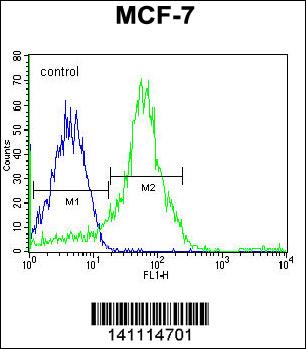
ESR2 Antibody (Center) flow cytometric analysis of MCF-7 cells (right histogram) compared to a negative control cell (left histogram).Alexa Fluor 488-conjugated donkey anti-rabbit lgG secondary antibodies were used for the analysis.Application
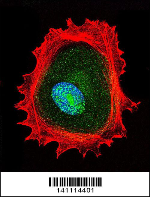
Confocal immunofluorescent analysis of ESR2 Antibody (Center) with MCF-7 cell followed by Alexa Fluor 488-conjugated goat anti-rabbit lgG (green). Actin filaments have been labeled with Alexa Fluor 555 phalloidin (red). DAPI was used to stain the cell nuclear (blue).Application
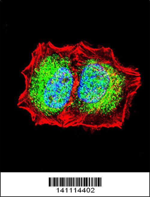
Confocal immunofluorescent analysis of ESR2 Antibody (Center) with MDA-MB453 cell followed by Alexa Fluor 488-conjugated goat anti-rabbit lgG (green). Actin filaments have been labeled with Alexa Fluor 555 phalloidin (red). DAPI was used to stain the cell nuclear (blue).Application
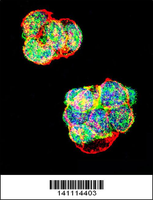
Confocal immunofluorescent analysis of ESR2 Antibody (Center) with T47D cell followed by Alexa Fluor 488-conjugated goat anti-rabbit lgG (green). Actin filaments have been labeled with Alexa Fluor 555 phalloidin (red). DAPI was used to stain the cell nuclear (blue).| Product Name | ESR2 Antibody (Center) |
|---|---|
| Antibody Type | Primary Antibodies |
| Clonality | Polyclonal |
|---|---|
| Isotype | Ig |
| Clone Number | RB41114 |
| Host Species | Rabbit |
| Tested Applications | WB,IHC,FC,IF |
| WB:1:2000 IHC |
|
| Species Reactivity | Human |
| Concentration | 1mg/ml |
| Gene Synonyms | ESTRB, NR3A2 |
|---|---|
| Alternative Names | ESR2 ESTRB NR3A2 Estrogen receptor beta Nuclear receptor subfamily 3 group A member 2 |
| Molecular Weight(MW) | 59216 Da |
| Function | Nuclear hormone receptor. Binds estrogens with an affinity similar to that of ESR1, and activates expression of reporter genes containing estrogen response elements (ERE) in an estrogen-dependent manner. Isoform beta-cx lacks ligand binding ability and has no or only very low ere binding activity resulting in the loss of ligand-dependent transactivation ability. DNA- binding by ESR1 and ESR2 is rapidly lost at 37 degrees Celsius in the absence of ligand while in the presence of 17 beta-estradiol and 4-hydroxy-tamoxifen loss in DNA-binding at elevated temperature is more gradual |
| Tissue Specificity | Isoform beta-1 is expressed in testis and ovary, and at a lower level in heart, brain, placenta, liver, skeletal muscle, spleen, thymus, prostate, colon, bone marrow, mammary gland and uterus. Also found in uterine bone, breast, and ovarian tumor cell lines, but not in colon and liver tumors Isoform beta-2 is expressed in spleen, thymus, testis and ovary and at a lower level in skeletal muscle, prostate, colon, small intestine, leukocytes, bone marrow, mammary gland and uterus Isoform beta-3 is found in testis. Isoform beta-4 is expressed in testis, and at a lower level in spleen, thymus, ovary, mammary gland and uterus. Isoform beta-5 is expressed in testis, placenta, skeletal muscle, spleen and leukocytes, and at a lower level in heart, lung, liver, kidney, pancreas, thymus, prostate, colon, small intestine, bone marrow, mammary gland and uterus. Not expressed in brain |
| Cellular Localization | Nucleus. |
| Entrez Gene | 2100 |
|---|---|
| Nucleotide Accession | NP_001035365.1 |
Application
ESR2 Antibody (Center) (Cat. #AX10006) western blot analysis in T47D,MDA-MB453,Hela cell line lysates (35ug/lane).This demonstrates the ESR2 antibody detected the ESR2 protein (arrow).
Application
ESR2 Antibody (Center) immunohistochemistry analysis in formalin fixed and paraffin embedded human testis tissue followed by peroxidase conjugation of the secondary antibody and DAB staining.This data demonstrates the use of ESR2 Antibody (Center) for immunohistochemistry. Clinical relevance has not been evaluated.
Application
ESR2 Antibody (Center) immunohistochemistry analysis in formalin fixed and paraffin embedded human ovarian carcinoma followed by peroxidase conjugation of the secondary antibody and DAB staining.This data demonstrates the use of ESR2 Antibody (Center) for immunohistochemistry. Clinical relevance has not been evaluated.
Application
ESR2 Antibody (Center) flow cytometric analysis of MCF-7 cells (right histogram) compared to a negative control cell (left histogram).Alexa Fluor 488-conjugated donkey anti-rabbit lgG secondary antibodies were used for the analysis.
Application
Confocal immunofluorescent analysis of ESR2 Antibody (Center) with MCF-7 cell followed by Alexa Fluor 488-conjugated goat anti-rabbit lgG (green). Actin filaments have been labeled with Alexa Fluor 555 phalloidin (red). DAPI was used to stain the cell nuclear (blue).
Application
Confocal immunofluorescent analysis of ESR2 Antibody (Center) with MDA-MB453 cell followed by Alexa Fluor 488-conjugated goat anti-rabbit lgG (green). Actin filaments have been labeled with Alexa Fluor 555 phalloidin (red). DAPI was used to stain the cell nuclear (blue).
Application
Confocal immunofluorescent analysis of ESR2 Antibody (Center) with T47D cell followed by Alexa Fluor 488-conjugated goat anti-rabbit lgG (green). Actin filaments have been labeled with Alexa Fluor 555 phalloidin (red). DAPI was used to stain the cell nuclear (blue).| Application Notes | WB:1:2000 IHC |
|---|
| Form | Liquid |
|---|---|
| Storage Instructions | For short-term storage, store at 4° C. For long-term storage, aliquot and store at -20ºC or below. Avoid multiple freeze-thaw cycles. |
| Storage Buffer | Purified polyclonal antibody supplied in PBS with 0.09% (W/V) sodium azide. This antibody is purified through a protein A column, followed by peptide affinity purification. |


