Application
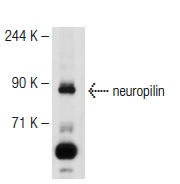
Figure 1: Western blot analysis using anti-FER mAb against NIH/3T3 (1), A549 (2) and SK-MEL-5 (3) cell lysate.Application
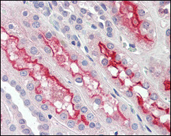
Figure 2: Immunohistochemical analysis of paraffin-embedded human kidney tissues using anti-FER mAb.Application
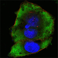
Figure 3: Confocal immunofluorescence analysis of Hela cells using anti-FER mAb (green). Red: Actin filaments have been labeled with Alexa Fluor-555 phalloidin. Blue: DRAQ5 fluorescent DNA dye.| Product Name | FER Antibody |
|---|---|
| Antibody Type | Primary Antibodies |
| Clonality | Monoclonal |
|---|---|
| Isotype | IgG1 |
| Clone Number | 5D2C4 |
| Host Species | Mouse |
| Tested Applications | WBIHCIF |
| WB:1:500 - 2000. IHC |
|
| Species Reactivity | Human |
| Concentration | 1mg/ml |
| Gene Synonyms | TYK3 |
|---|---|
| Alternative Names | TYK3 FER |
| Molecular Weight(MW) | 94638 Da |
| Function | Tyrosine-protein kinase that acts downstream of cell surface receptors for growth factors and plays a role in the regulation of the actin cytoskeleton, microtubule assembly, lamellipodia formation, cell adhesion, cell migration and chemotaxis. Acts downstream of EGFR, KIT, PDGFRA and PDGFRB. Acts downstream of EGFR to promote activation of NF-kappa-B and cell proliferation. May play a role in the regulation of the mitotic cell cycle. Plays a role in the insulin receptor signaling pathway and in activation of phosphatidylinositol 3-kinase. Acts downstream of the activated FCER1 receptor and plays a role in FCER1 (high affinity immunoglobulin epsilon receptor)-mediated signaling in mast cells. Plays a role in the regulation of mast cell degranulation. Plays a role in leukocyte recruitment and diapedesis in response to bacterial lipopolysaccharide (LPS) Plays a role in synapse organization, trafficking of synaptic vesicles, the generation of excitatory postsynaptic currents and neuron-neuron synaptic transmission. Plays a role in neuronal cell death after brain damage. Phosphorylates CTTN, CTNND1, PTK2/FAK1, GAB1, PECAM1 and PTPN11. May phosphorylate JUP and PTPN1. Can phosphorylate STAT3, but the biological relevance of this depends on cell type and stimulus |
| Tissue Specificity | Isoform 1 is detected in normal colon and in fibroblasts (at protein level). Isoform 3 is detected in normal testis, in colon carcinoma-derived metastases in lung, liver and ovary, and in colon carcinoma and hepato carcinoma cell lines (at protein level). Isoform 3 is not detected in normal colon or in normal fibroblasts (at protein level). Widely expressed |
| Cellular Localization | Cytoplasm. Cytoplasm, cytoskeleton. Cell membrane; Peripheral membrane protein; Cytoplasmic side. Cell projection. Cell junction. Membrane; Peripheral membrane protein; Cytoplasmic side. Nucleus. Cytoplasm, cell cortex. Note=Associated with the chromatin. Detected on microtubules in polarized and motile vascular endothelial cells. Colocalizes with F-actin at the cell cortex. Colocalizes with PECAM1 and CTNND1 at nascent cell- cell contacts |
| Entrez Gene | 2241 |
|---|---|
| Nucleotide Accession | NP_005237.2 |
Application
Figure 1: Western blot analysis using anti-FER mAb against NIH/3T3 (1), A549 (2) and SK-MEL-5 (3) cell lysate.
Application
Figure 2: Immunohistochemical analysis of paraffin-embedded human kidney tissues using anti-FER mAb.
Application
Figure 3: Confocal immunofluorescence analysis of Hela cells using anti-FER mAb (green). Red: Actin filaments have been labeled with Alexa Fluor-555 phalloidin. Blue: DRAQ5 fluorescent DNA dye.| Application Notes | WB:1:500 - 2000. IHC |
|---|
| Form | Liquid |
|---|---|
| Storage Instructions | For short-term storage, store at 4° C. For long-term storage, aliquot and store at -20ºC or below. Avoid multiple freeze-thaw cycles. |
| Storage Buffer | Purified recombinant fragment of human FER expressed in E. Coli. |


