Application
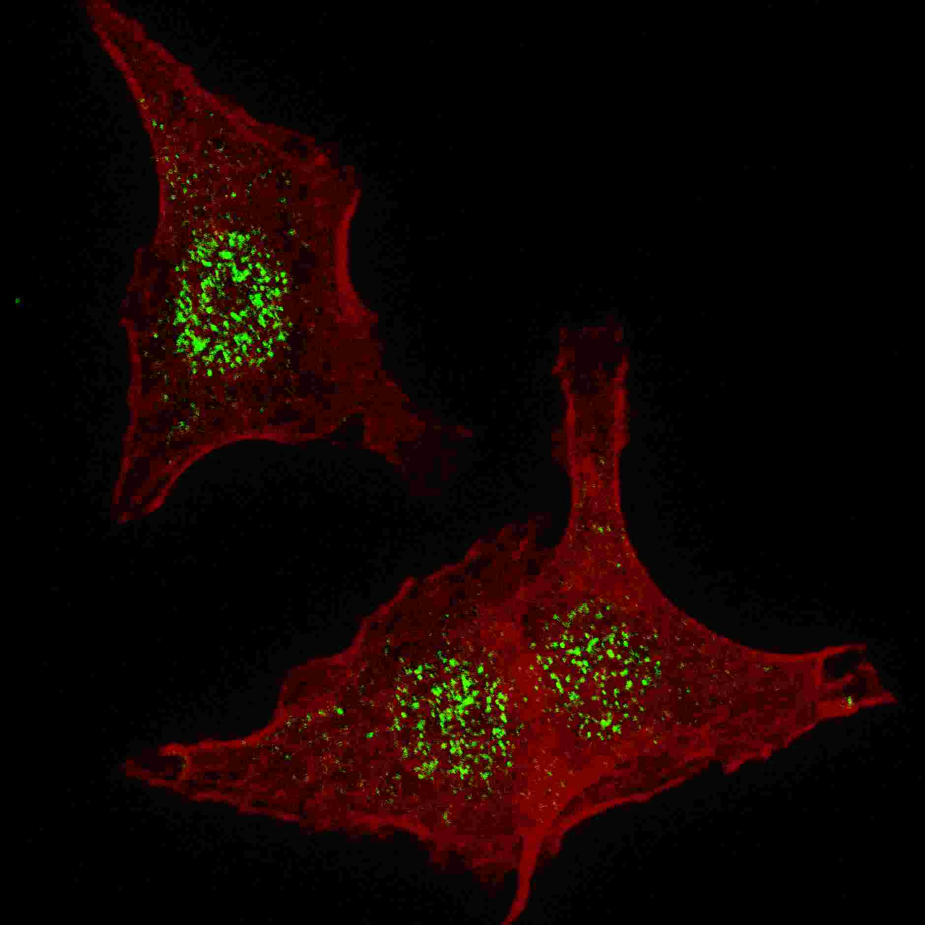
Fluorescent confocal image of HeLa cells stained with FGFR4 (N-term) antibody. HeLa cells were fixed with 4% PFA (20 min), permeabilized with Triton X-100 (0.2%, 30 min). Cells were then incubated with AP7639a FGFR4 (N-term) primary antibody (1:200, 2 h at room temperature). For secondary antibody, Alexa Fluor® 488 conjugated donkey anti-rabbit antibody (green) was used (1:1000, 1h). Nuclei were counterstained with Hoechst 33342 (blue) (10 μg/ml, 5 min). Note the highly specific localization of the FGFR4 mainly to the nucleus, supported by Human Protein Atlas Data (http://www.proteinatlas.org/ENSG00000160867).Application
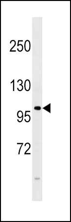
FGFR4 Antibody (E39) western blot analysis in 293 cell line lysates (35ug/lane).This demonstrates the FGFR4 antibody detected the FGFR4 protein (arrow).Application
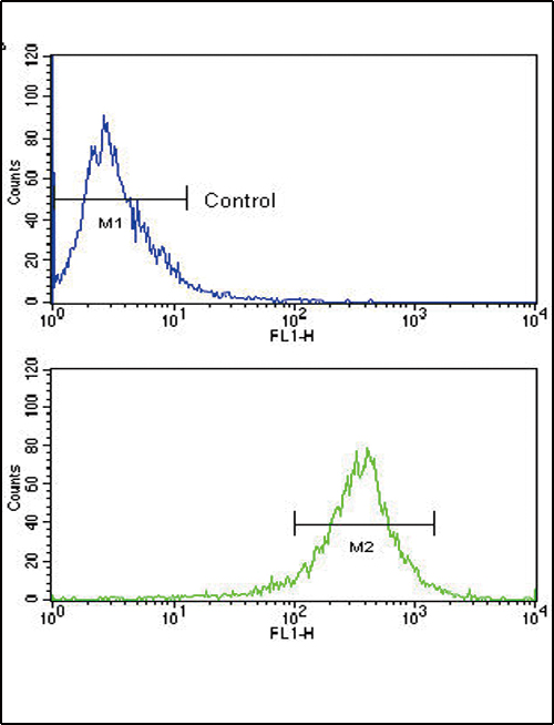
Flow cytometric analysis of WiDr cells using FGFR4 Antibody (N-term) (bottom histogram) compared to a negative control cell (top histogram). FITC-conjugated goat-anti-rabbit secondary antibodies were used for the analysis.| Product Name | FGFR4 Antibody (N-term) |
|---|---|
| Antibody Type | Primary Antibodies |
| Clonality | Polyclonal |
|---|---|
| Isotype | Ig |
| Host Species | Rabbit |
| Tested Applications | IFWBFC |
| IF:1:200 WB |
|
| Species Reactivity | Human |
| Concentration | 1mg/ml |
| Gene Synonyms | JTK2, TKF |
|---|---|
| Alternative Names | FGFR4 JTK2 TKF Fibroblast growth factor receptor 4 CD_antigen=CD334 Flags: Precursor |
| Molecular Weight(MW) | 87954 Da |
| Function | Tyrosine-protein kinase that acts as cell-surface receptor for fibroblast growth factors and plays a role in the regulation of cell proliferation, differentiation and migration, and in regulation of lipid metabolism, bile acid biosynthesis, glucose uptake, vitamin D metabolism and phosphate homeostasis Required for normal down-regulation of the expression of CYP7A1, the rate-limiting enzyme in bile acid synthesis, in response to FGF19. Phosphorylates PLCG1 and FRS2. Ligand binding leads to the activation of several signaling cascades. Activation of PLCG1 leads to the production of the cellular signaling molecules diacylglycerol and inositol 1,4,5-trisphosphate. Phosphorylation of FRS2 triggers recruitment of GRB2, GAB1, PIK3R1 and SOS1, and mediates activation of RAS, MAPK1/ERK2, MAPK3/ERK1 and the MAP kinase signaling pathway, as well as of the AKT1 signaling pathway. Promotes SRC-dependent phosphorylation of the matrix protease MMP14 and its lysosomal degradation. FGFR4 signaling is down-regulated by receptor internalization and degradation; MMP14 promotes internalization and degradation of FGFR4. Mutations that lead to constitutive kinase activation or impair normal FGFR4 inactivation lead to aberrant signaling |
| Tissue Specificity | Expressed in gastrointestinal epithelial cells, pancreas, and gastric and pancreatic cancer cell lines |
| Cellular Localization | Cell membrane; Single-pass type I membrane protein. Endosome. Endoplasmic reticulum. Note=Internalized from the cell membrane to recycling endosomes, and from there back to the cell membrane |
| Entrez Gene | 2264 |
|---|
Application
Fluorescent confocal image of HeLa cells stained with FGFR4 (N-term) antibody. HeLa cells were fixed with 4% PFA (20 min), permeabilized with Triton X-100 (0.2%, 30 min). Cells were then incubated with AP7639a FGFR4 (N-term) primary antibody (1:200, 2 h at room temperature). For secondary antibody, Alexa Fluor® 488 conjugated donkey anti-rabbit antibody (green) was used (1:1000, 1h). Nuclei were counterstained with Hoechst 33342 (blue) (10 μg/ml, 5 min). Note the highly specific localization of the FGFR4 mainly to the nucleus, supported by Human Protein Atlas Data (http://www.proteinatlas.org/ENSG00000160867).
Application
FGFR4 Antibody (E39) western blot analysis in 293 cell line lysates (35ug/lane).This demonstrates the FGFR4 antibody detected the FGFR4 protein (arrow).
Application
Flow cytometric analysis of WiDr cells using FGFR4 Antibody (N-term) (bottom histogram) compared to a negative control cell (top histogram). FITC-conjugated goat-anti-rabbit secondary antibodies were used for the analysis.| Application Notes | IF:1:200 WB |
|---|
| Form | Liquid |
|---|---|
| Storage Instructions | For short-term storage, store at 4° C. For long-term storage, aliquot and store at -20ºC or below. Avoid multiple freeze-thaw cycles. |
| Storage Buffer | Purified polyclonal antibody supplied in PBS with 0.09% (W/V) sodium azide. This antibody is purified through a protein G column, eluted with high and low pH buffers and neutralized immediately, followed by dialysis against PBS. |


