Application
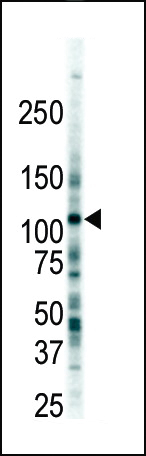
Western blot analysis of anti-FLT3 Pab in 293 cell lysate. FLT3 (arrow) was detected using purified Pab. Secondary HRP-anti-rabbit was used for signal visualization with chemiluminescence.| Product Name | FLT3 (CD135) Antibody (C-term) |
|---|---|
| Antibody Type | Primary Antibodies |
| Antigen Alias | FLT3; CD135; FLK2; STK1; Receptor-type tyrosine-protein kinase FLT3; FL cytokine receptor; Fetal liver kinase-2; Fms-like tyrosine kinase 3; Stem cell tyrosine kinase 1; CD_antigen=CD135; Flags: Precursor |
| Clonality | Polyclonal |
|---|---|
| Isotype | Ig |
| Host Species | Rabbit |
| Tested Applications | WB |
| WB:1:100~500 |
|
| Species Reactivity | Human |
| Concentration | 1mg/ml |
| Gene Synonyms | CD135 FLK2 STK1 |
|---|---|
| Alternative Names | FLT3 CD135 FLK2 STK1 Receptor-type tyrosine-protein kinase FLT3 FL cytokine receptor Fetal liver kinase-2 Fms-like tyrosine kinase 3 Stem cell tyrosine kinase 1 CD_antigen=CD135 Flags: Precursor |
| Molecular Weight(MW) | 112903 Da |
| Function | Tyrosine-protein kinase that acts as cell-surface receptor for the cytokine FLT3LG and regulates differentiation, proliferation and survival of hematopoietic progenitor cells and of dendritic cells. Promotes phosphorylation of SHC1 and AKT1, and activation of the downstream effector MTOR. Promotes activation of RAS signaling and phosphorylation of downstream kinases, including MAPK1/ERK2 and/or MAPK3/ERK1. Promotes phosphorylation of FES, FER, PTPN6/SHP, PTPN11/SHP-2, PLCG1, and STAT5A and/or STAT5B Activation of wild-type FLT3 causes only marginal activation of STAT5A or STAT5B. Mutations that cause constitutive kinase activity promote cell proliferation and resistance to apoptosis via the activation of multiple signaling pathways |
| Tissue Specificity | Detected in bone marrow, in hematopoietic stem cells, in myeloid progenitor cells and in granulocyte/macrophage progenitor cells (at protein level). Detected in bone marrow, liver, thymus, spleen and lymph node, and at low levels in kidney and pancreas. Highly expressed in T-cell leukemia |
| Cellular Localization | Membrane; Single-pass type I membrane protein. Endoplasmic reticulum lumen. Note=Constitutively activated mutant forms with internal tandem duplications are less efficiently transported to the cell surface and a significant proportion is retained in an immature form in the endoplasmic reticulum lumen. The activated kinase is rapidly targeted for degradation |
| Entrez Gene | 2322 |
|---|
Application
Western blot analysis of anti-FLT3 Pab in 293 cell lysate. FLT3 (arrow) was detected using purified Pab. Secondary HRP-anti-rabbit was used for signal visualization with chemiluminescence.| Application Notes | WB:1:100~500 |
|---|
| Form | Liquid |
|---|---|
| Storage Instructions | For short-term storage, store at 4° C. For long-term storage, aliquot and store at -20ºC or below. Avoid multiple freeze-thaw cycles. |
| Storage Buffer | Purified polyclonal antibody supplied in PBS with 0.09% (W/V) sodium azide. This antibody is purified through a protein G column, eluted with high and low pH buffers and neutralized immediately, followed by dialysis against PBS. |


