Application
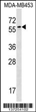
FSD1 Antibody (N-term) western blot analysis in MDA-MB453 cell line lysates (35ug/lane).This demonstrates the FSD1 antibody detected the FSD1 protein (arrow).| Product Name | FSD1 Antibody (N-term) |
|---|---|
| Antibody Type | Primary Antibodies |
| Antigen Alias | FSD1; GLFND; MIR1; Fibronectin type III and SPRY domain-containing protein 1; MID1-related protein 1; Microtubule-associated protein GLFND |
| Clonality | Polyclonal |
|---|---|
| Isotype | Ig |
| Host Species | Rabbit |
| Tested Applications | WB |
| WB:1:100~500 |
|
| Species Reactivity | Human |
| Concentration | 1mg/ml |
| Gene Synonyms | GLFND MIR1 |
|---|---|
| Alternative Names | FSD1 GLFND MIR1 Fibronectin type III and SPRY domain-containing protein 1 MID1-related protein 1 Microtubule-associated protein GLFND |
| Molecular Weight(MW) | 55820 Da |
| Function | May be involved in microtubule organization and stabilization |
| Tissue Specificity | Highly expressed in brain tissues, including cerebellum, cerebral cortex, medulla, occipital pole, frontal lobe, temporal lobe and putamen. Lower expression in spinal chord |
| Cellular Localization | Cytoplasm, cytoskeleton, centrosome. Nucleus. Cytoplasm. Cleavage furrow. Note=Cell-cycle-dependent association with the centrosome. Colocalizes with a subpopulation of microtubules. Does not associates with microtubules during mitosis but reassociates with microtubules during cytokinesis Localizes to the central portions of a small subset of microtubules in interphase cells and a subpopulation of microtubules in the cleavage furrow, not present in the mitotic spindle |
| Entrez Gene | 79187 |
|---|---|
| Nucleotide Accession | NP_077309.1 |
Application
FSD1 Antibody (N-term) western blot analysis in MDA-MB453 cell line lysates (35ug/lane).This demonstrates the FSD1 antibody detected the FSD1 protein (arrow).| Application Notes | WB:1:100~500 |
|---|
| Form | Liquid |
|---|---|
| Storage Instructions | For short-term storage, store at 4° C. For long-term storage, aliquot and store at -20ºC or below. Avoid multiple freeze-thaw cycles. |
| Storage Buffer | Purified polyclonal antibody supplied in PBS with 0.09% (W/V) sodium azide. This antibody is purified through a protein A column, followed by peptide affinity purification. |


