Application
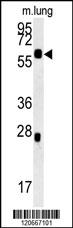
HCK Antibody (N-term) western blot analysis in mouse lung tissue lysates (15ug/lane).This demonstrates the HCK antibody detected HCK protein (arrow).Application
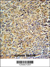
HCK Antibody (N-term) immunohistochemistry analysis in formalin fixed and paraffin embedded human spleen tissue followed by peroxidase conjugation of the secondary antibody and DAB staining. This data demonstrates the use of the HCK Antibody (N-term) for immunohistochemistry. Clinical relevance has not been evaluated.Application
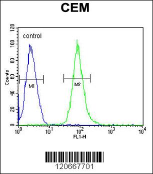
HCK Antibody (N-term) flow cytometric analysis of CEM cells (right histogram) compared to a negative control cell (left histogram).FITC-conjugated goat-anti-rabbit secondary antibodies were used for the analysis.| Product Name | HCK Antibody (N-term) |
|---|---|
| Antibody Type | Primary Antibodies |
| Antigen Alias | HCK; Tyrosine-protein kinase HCK; Hematopoietic cell kinase; Hemopoietic cell kinase; p59-HCK/p60-HCK; p59Hck; p61Hck |
| Clonality | Polyclonal |
|---|---|
| Isotype | Ig |
| Host Species | Rabbit |
| Tested Applications | WBIHCFC |
| WB:1:100~500 IHC |
|
| Species Reactivity | Human |
| Concentration | 1mg/ml |
| Alternative Names | HCK Tyrosine-protein kinase HCK Hematopoietic cell kinase Hemopoietic cell kinase p59-HCK/p60-HCK p59Hck p61Hck |
|---|---|
| Molecular Weight(MW) | 59600 Da |
| Function | Non-receptor tyrosine-protein kinase found in hematopoietic cells that transmits signals from cell surface receptors and plays an important role in the regulation of innate immune responses, including neutrophil, monocyte, macrophage and mast cell functions, phagocytosis, cell survival and proliferation, cell adhesion and migration. Acts downstream of receptors that bind the Fc region of immunoglobulins, such as FCGR1A and FCGR2A, but also CSF3R, PLAUR, the receptors for IFNG, IL2, IL6 and IL8, and integrins, such as ITGB1 and ITGB2. During the phagocytic process, mediates mobilization of secretory lysosomes, degranulation, and activation of NADPH oxidase to bring about the respiratory burst. Plays a role in the release of inflammatory molecules. Promotes reorganization of the actin cytoskeleton and actin polymerization, formation of podosomes and cell protrusions. Inhibits TP73-mediated transcription activation and TP73-mediated apoptosis. Phosphorylates CBL in response to activation of immunoglobulin gamma Fc region receptors Phosphorylates ADAM15, BCR, ELMO1, FCGR2A, GAB1, GAB2, RAPGEF1, STAT5B, TP73, VAV1 and WAS |
| Tissue Specificity | Detected in monocytes and neutrophils (at protein level). Expressed predominantly in cells of the myeloid and B-lymphoid lineages. Highly expressed in granulocytes Detected in tonsil |
| Cellular Localization | Isoform 1: Lysosome. Membrane; Lipid-anchor. Cell projection, podosome membrane; Lipid-anchor. Cytoplasm, cytosol. Note=Associated with specialized secretory lysosomes called azurophil granules. At least half of this isoform is found in the cytoplasm, some of this fraction is myristoylated Cytoplasmic vesicle, secretory vesicle. Cytoplasm, cytosol |
Application
HCK Antibody (N-term) western blot analysis in mouse lung tissue lysates (15ug/lane).This demonstrates the HCK antibody detected HCK protein (arrow).
Application
HCK Antibody (N-term) immunohistochemistry analysis in formalin fixed and paraffin embedded human spleen tissue followed by peroxidase conjugation of the secondary antibody and DAB staining. This data demonstrates the use of the HCK Antibody (N-term) for immunohistochemistry. Clinical relevance has not been evaluated.
Application
HCK Antibody (N-term) flow cytometric analysis of CEM cells (right histogram) compared to a negative control cell (left histogram).FITC-conjugated goat-anti-rabbit secondary antibodies were used for the analysis.| Application Notes | WB:1:100~500 IHC |
|---|
| Form | Liquid |
|---|---|
| Storage Instructions | For short-term storage, store at 4° C. For long-term storage, aliquot and store at -20ºC or below. Avoid multiple freeze-thaw cycles. |
| Storage Buffer | Purified polyclonal antibody supplied in PBS with 0.09% (W/V) sodium azide. This antibody is purified through a protein A column, followed by peptide affinity purification. |


