Application
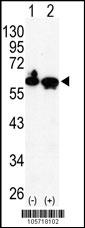
Western blot analysis of HDAC2 (arrow) using rabbit polyclonal HDAC2 Antibody (C-term) .293 cell lysates (2 ug/lane) either nontransfected (Lane 1) or transiently transfected with the HDAC2 gene (Lane 2) (Origene Technologies).Application
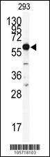
Western blot analysis of anti-HDAC2 Antibody (C-term) in 293 cell line lysates (35ug/lane). HDAC2(arrow) was detected using the purified Pab.Application
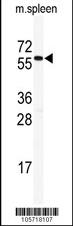
Western blot analysis of anti-HDAC2 Antibody (C-term) in mouse spleen tissue lysates (35ug/lane). HDAC2(arrow) was detected using the purified Pab.Application
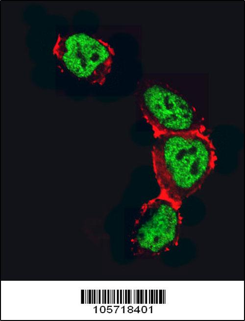
Confocal immunofluorescent analysis of HDAC2 Antibody (C-term)(Cat#AP1102b) with 293 cell followed by Alexa Fluor 488-conjugated goat anti-rabbit lgG (green).Actin filaments have been labeled with Alexa Fluor 555 phalloidin (red).Application
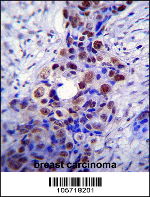
HDAC2 Antibody (C-term) immunohistochemistry analysis in formalin fixed and paraffin embedded human breast carcinoma followed by peroxidase conjugation of the secondary antibody and DAB staining.This data demonstrates the use of HDAC2 Antibody (C-term) for immunohistochemistry. Clinical relevance has not been evaluated.Application
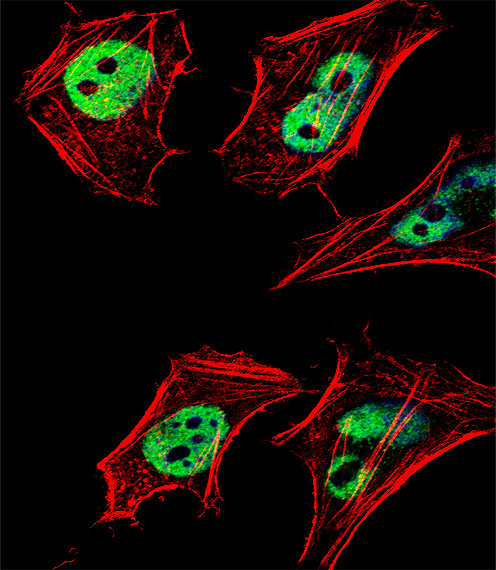
Fluorescent confocal image of Hela cell stained with HDAC2 Antibody (C-term)(Cat#AP1102b).Hela cells were fixed with 4% PFA (20 min), permeabilized with Triton X-100 (0.1%, 10 min), then incubated with HDAC2 primary antibody (1:25, 1 h at 37℃). For secondary antibody, Alexa Fluor® 488 conjugated donkey anti-rabbit antibody (green) was used (1:400, 50 min at 37℃).Cytoplasmic actin was counterstained with Alexa Fluor® 555 (red) conjugated Phalloidin (7units/ml, 1 h at 37℃). Nuclei were counterstained with DAPI (blue) (10 µg/ml, 10 min). hHDAC2 immunoreactivity is localized to Nucleus significantly.| Product Name | HDAC2 Antibody (C-term) |
|---|---|
| Antibody Type | Primary Antibodies |
| Antigen Alias | HDAC2; Histone deacetylase 2 |
| Clonality | Polyclonal |
|---|---|
| Isotype | Ig |
| Host Species | Rabbit |
| Tested Applications | WBIFIHC |
| WB:1:100~500 IF |
|
| Species Reactivity | Human |
| Concentration | 1mg/ml |
| Alternative Names | HDAC2 Histone deacetylase 2 |
|---|---|
| Molecular Weight(MW) | 55364 Da |
| Function | Responsible for the deacetylation of lysine residues on the N-terminal part of the core histones (H2A, H2B, H3 and H4) Histone deacetylation gives a tag for epigenetic repression and plays an important role in transcriptional regulation, cell cycle progression and developmental events. Histone deacetylases act via the formation of large multiprotein complexes. Forms transcriptional repressor complexes by associating with MAD, SIN3, YY1 and N-COR. Interacts in the late S-phase of DNA-replication with DNMT1 in the other transcriptional repressor complex composed of DNMT1, DMAP1, PCNA, CAF1. Deacetylates TSHZ3 and regulates its transcriptional repressor activity. Component of a RCOR/GFI/KDM1A/HDAC complex that suppresses, via histone deacetylase (HDAC) recruitment, a number of genes implicated in multilineage blood cell development |
| Tissue Specificity | Widely expressed; lower levels in brain and lung |
| Cellular Localization | Nucleus. |
| Entrez Gene | 3066 |
|---|
Application
Western blot analysis of HDAC2 (arrow) using rabbit polyclonal HDAC2 Antibody (C-term) .293 cell lysates (2 ug/lane) either nontransfected (Lane 1) or transiently transfected with the HDAC2 gene (Lane 2) (Origene Technologies).
Application
Western blot analysis of anti-HDAC2 Antibody (C-term) in 293 cell line lysates (35ug/lane). HDAC2(arrow) was detected using the purified Pab.
Application
Western blot analysis of anti-HDAC2 Antibody (C-term) in mouse spleen tissue lysates (35ug/lane). HDAC2(arrow) was detected using the purified Pab.
Application
Confocal immunofluorescent analysis of HDAC2 Antibody (C-term)(Cat#AP1102b) with 293 cell followed by Alexa Fluor 488-conjugated goat anti-rabbit lgG (green).Actin filaments have been labeled with Alexa Fluor 555 phalloidin (red).
Application
HDAC2 Antibody (C-term) immunohistochemistry analysis in formalin fixed and paraffin embedded human breast carcinoma followed by peroxidase conjugation of the secondary antibody and DAB staining.This data demonstrates the use of HDAC2 Antibody (C-term) for immunohistochemistry. Clinical relevance has not been evaluated.
Application
Fluorescent confocal image of Hela cell stained with HDAC2 Antibody (C-term)(Cat#AP1102b).Hela cells were fixed with 4% PFA (20 min), permeabilized with Triton X-100 (0.1%, 10 min), then incubated with HDAC2 primary antibody (1:25, 1 h at 37℃). For secondary antibody, Alexa Fluor® 488 conjugated donkey anti-rabbit antibody (green) was used (1:400, 50 min at 37℃).Cytoplasmic actin was counterstained with Alexa Fluor® 555 (red) conjugated Phalloidin (7units/ml, 1 h at 37℃). Nuclei were counterstained with DAPI (blue) (10 µg/ml, 10 min). hHDAC2 immunoreactivity is localized to Nucleus significantly.| Application Notes | WB:1:100~500 IF |
|---|
| Form | Liquid |
|---|---|
| Storage Instructions | For short-term storage, store at 4° C. For long-term storage, aliquot and store at -20ºC or below. Avoid multiple freeze-thaw cycles. |
| Storage Buffer | Purified polyclonal antibody supplied in PBS with 0.09% (W/V) sodium azide. This antibody is purified through a protein G column, eluted with high and low pH buffers and neutralized immediately, followed by dialysis against PBS. |


