Application
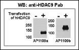
Both anti-HDAC9 N-term (AP1109a) and C-term (AP1109b) Pab were tested by WB and IP-WB using HeLa and HeLa-HDAC9 transfected cells. Top figure shows both Pab specifically detect HDAC9 in HeLa-HDAC9 transfected cell but not HeLa alone.Application
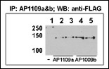
This figure shows that both Pab can immunoprecipitate (IP) HDAC9 from HeLa-HDAC9 tranfected cells. (Data kindly provided by Dr. Zhigang Yuan, H. Lee Moffitt Cancer Center and Research Institute, Tampa, FL).Application
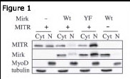
Figure 1: Immunoblots for MITR (AP1109a HDAC9 N-term antibody), Mirk, MyoD and tubulin proteins are shown for cytoplasmic (Cyt) and nuclear (N) extracts from undifferentiated C2C12 myoblasts. Before cell collection for fractionation, the cells are transfected with plasmids coding for Mirk (Wt), kinase-inactive Mirk (YF) or MITR. Data courtesy of laboratory of Dr. Eileen Friedman. Dept of Pathology, Upstate Medical University, State University of New York.Application
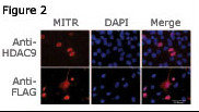
Figure 2: Immunofluorescence staining of MITR for a compartmentalization study in undifferentiated C2C12 myoblasts transfected with a MITR-expressing plasmid. MITR is detected by using the HDAC9 N-term antibody (top panel) or a FLAG antibody (bottom panel) detecting a FLAG epitope fused at the N-term end of the MITR construct. Data courtesy of laboratory of Dr. Eileen Friedman. Dept of Pathology, Upstate Medical University, State University of New York.Application
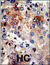
Formalin-fixed and paraffin-embedded human cancer tissue reacted with the primary antibody, which was peroxidase-conjugated to the secondary antibody, followed by AEC staining. This data demonstrates the use of this antibody for immunohistochemistry; clinical relevance has not been evaluated. BC = breast carcinoma; HC = hepatocarcinoma.| Product Name | HDAC9 Antibody (N-term) |
|---|---|
| Antibody Type | Primary Antibodies |
| Clonality | Polyclonal |
|---|---|
| Isotype | Ig |
| Host Species | Rabbit |
| Tested Applications | WB,IP,IF,IHC |
| WB:1:1000 IP |
|
| Species Reactivity | Human |
| Concentration | 1mg/ml |
| Gene Synonyms | HDAC7, HDAC7B, HDRP, KIAA0744, MITR |
|---|---|
| Alternative Names | HDAC9 HDAC7 HDAC7B HDRP KIAA0744 MITR Histone deacetylase 9 Histone deacetylase 7B Histone deacetylase-related protein MEF2-interacting transcription repressor MITR |
| Molecular Weight(MW) | 111297 Da |
| Function | Responsible for the deacetylation of lysine residues on the N-terminal part of the core histones (H2A, H2B, H3 and H4) Histone deacetylation gives a tag for epigenetic repression and plays an important role in transcriptional regulation, cell cycle progression and developmental events. Represses MEF2-dependent transcription |
| Tissue Specificity | Broadly expressed, with highest levels in brain, heart, muscle and testis. Isoform 3 is present in human bladder carcinoma cells (at protein level) |
| Cellular Localization | Nucleus (By similarity). |
| Entrez Gene | 9734 |
|---|
Application
Both anti-HDAC9 N-term (AP1109a) and C-term (AP1109b) Pab were tested by WB and IP-WB using HeLa and HeLa-HDAC9 transfected cells. Top figure shows both Pab specifically detect HDAC9 in HeLa-HDAC9 transfected cell but not HeLa alone.
Application
This figure shows that both Pab can immunoprecipitate (IP) HDAC9 from HeLa-HDAC9 tranfected cells. (Data kindly provided by Dr. Zhigang Yuan, H. Lee Moffitt Cancer Center and Research Institute, Tampa, FL).
Application
Figure 1: Immunoblots for MITR (AP1109a HDAC9 N-term antibody), Mirk, MyoD and tubulin proteins are shown for cytoplasmic (Cyt) and nuclear (N) extracts from undifferentiated C2C12 myoblasts. Before cell collection for fractionation, the cells are transfected with plasmids coding for Mirk (Wt), kinase-inactive Mirk (YF) or MITR. Data courtesy of laboratory of Dr. Eileen Friedman. Dept of Pathology, Upstate Medical University, State University of New York.
Application
Figure 2: Immunofluorescence staining of MITR for a compartmentalization study in undifferentiated C2C12 myoblasts transfected with a MITR-expressing plasmid. MITR is detected by using the HDAC9 N-term antibody (top panel) or a FLAG antibody (bottom panel) detecting a FLAG epitope fused at the N-term end of the MITR construct. Data courtesy of laboratory of Dr. Eileen Friedman. Dept of Pathology, Upstate Medical University, State University of New York.
Application
Formalin-fixed and paraffin-embedded human cancer tissue reacted with the primary antibody, which was peroxidase-conjugated to the secondary antibody, followed by AEC staining. This data demonstrates the use of this antibody for immunohistochemistry; clinical relevance has not been evaluated. BC = breast carcinoma; HC = hepatocarcinoma.| Application Notes | WB:1:1000 IP |
|---|
| Form | Liquid |
|---|---|
| Storage Instructions | For short-term storage, store at 4° C. For long-term storage, aliquot and store at -20ºC or below. Avoid multiple freeze-thaw cycles. |
| Storage Buffer | Purified polyclonal antibody supplied in PBS with 0.09% (W/V) sodium azide. This antibody is purified through a protein G column, eluted with high and low pH buffers and neutralized immediately, followed by dialysis against PBS. |


