Application
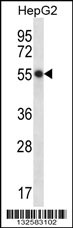
HEPACAM Antibody (C-term) western blot analysis in HepG2 cell line lysates (35ug/lane).This demonstrates the HEPACAM antibody detected the HEPACAM protein (arrow).| Product Name | HEPACAM Antibody (C-term) |
|---|---|
| Antibody Type | Primary Antibodies |
| Antigen Alias | HEPACAM; Hepatocyte cell adhesion molecule |
| Clonality | Polyclonal |
|---|---|
| Isotype | Ig |
| Host Species | Rabbit |
| Tested Applications | WB |
| WB:1:100~500 |
|
| Species Reactivity | Human |
| Concentration | 1mg/ml |
| Alternative Names | HEPACAM Hepatocyte cell adhesion molecule |
|---|---|
| Molecular Weight(MW) | 46026 Da |
| Function | Involved in regulating cell motility and cell-matrix interactions. May inhibit cell growth through suppression of cell proliferation |
| Tissue Specificity | This HEPACAM antibody is generated from rabbits immunized with a KLH conjugated synthetic peptide between 305-335 amino acids from the C-terminal region of human HEPACAM. |
| Cellular Localization | Cytoplasm. Membrane; Single-pass type I membrane protein; Cytoplasmic side. Note=In MCF-7 breast carcinoma and hepatic Hep 3B2.1-7 and Hep-G2 cell lines, localization of HEPACAM is cell density-dependent. In well spread cells, localized to punctate structures in the perinuclear membrane, cytoplasm, and at cell surface of protusions. In confluent cells, localized predominantly to the cytoplasmic membrane, particularly in areas of cell-cell contacts. Colocalizes with CDH1 |
| Entrez Gene | 220296 |
|---|---|
| Nucleotide Accession | NP_689935.2 |
Application
HEPACAM Antibody (C-term) western blot analysis in HepG2 cell line lysates (35ug/lane).This demonstrates the HEPACAM antibody detected the HEPACAM protein (arrow).| Application Notes | WB:1:100~500 |
|---|
| Form | Liquid |
|---|---|
| Storage Instructions | For short-term storage, store at 4° C. For long-term storage, aliquot and store at -20ºC or below. Avoid multiple freeze-thaw cycles. |
| Storage Buffer | Purified polyclonal antibody supplied in PBS with 0.09% (W/V) sodium azide. This antibody is purified through a protein A column, followed by peptide affinity purification. |


