Application
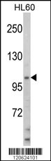
Western blot analysis of HIP116A Antibody (Center) in HL60 cell line lysates (35ug/lane). HIP116A (arrow) was detected using the purified Pab.Application
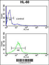
HIP116A Antibody (Center) flow cytometry analysis of HL-60 cells (bottom histogram) compared to a negative control cell (top histogram).FITC-conjugated goat-anti-rabbit secondary antibodies were used for the analysis.| Product Name | HIP116A Antibody (Center) |
|---|---|
| Antibody Type | Primary Antibodies |
| Clonality | Polyclonal |
|---|---|
| Isotype | Ig |
| Host Species | Rabbit |
| Tested Applications | WBFC |
| WB:1:50~100 FC |
|
| Species Reactivity | Human |
| Concentration | 1mg/ml |
| Gene Synonyms | HIP116A, RNF80, SMARCA3, SNF2L3, ZBU1 |
|---|---|
| Alternative Names | HLTF HIP116A RNF80 SMARCA3 SNF2L3 ZBU1 Helicase-like transcription factor DNA-binding protein/plasminogen activator inhibitor 1 regulator HIP116 RING finger protein 80 SWI/SNF-related matrix-associated actin-dependent regulator of chromatin subfa |
| Molecular Weight(MW) | 113929 Da |
| Function | Has both helicasee and E3 ubiquitin ligase activities. Possesses intrinsic ATP-dependent nucleosome-remodeling activity; This activity may be required for transcriptional activation or repression of specific target promoters (By similarity). These may include the SERPINE1 and HIV-1 promoters and the SV40 enhancer, to which this protein can bind directly. Plays a role in error-free postreplication repair (PRR) of damaged DNA and maintains genomic stability through acting as a ubiquitin ligase for 'Lys-63'-linked polyubiquitination of chromatin-bound PCNA |
| Tissue Specificity | Expressed in brain, heart, kidney, liver, lung, pancreas, placenta and skeletal muscle |
| Cellular Localization | Nucleus. |
| Entrez Gene | 6596 |
|---|
Application
Western blot analysis of HIP116A Antibody (Center) in HL60 cell line lysates (35ug/lane). HIP116A (arrow) was detected using the purified Pab.
Application
HIP116A Antibody (Center) flow cytometry analysis of HL-60 cells (bottom histogram) compared to a negative control cell (top histogram).FITC-conjugated goat-anti-rabbit secondary antibodies were used for the analysis.| Application Notes | WB:1:50~100 FC |
|---|
| Form | Liquid |
|---|---|
| Storage Instructions | For short-term storage, store at 4° C. For long-term storage, aliquot and store at -20ºC or below. Avoid multiple freeze-thaw cycles. |
| Storage Buffer | Purified polyclonal antibody supplied in PBS with 0.09% (W/V) sodium azide. This antibody is prepared by Saturated Ammonium Sulfate (SAS) precipitation followed by dialysis against PBS. |


