Application
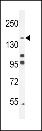
His6-DEP-1 western blot analysis in K562 cell line lysates (35ug/lane).This demonstrates the DEP-1 antibody detected the DEP-1 protein (arrow).Application
His6-DEP-1 Antibody immunohistochemistry analysis in formalin fixed and paraffin embedded human tonsil tissue followed by peroxidase conjugation of the secondary antibody and DAB staining.This data demonstrates the use of His6-DEP-1 Antibody for immunohistochemistry. Clinical relevance has not been evaluated.Application
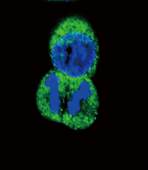
Confocal immunofluorescent analysis of His6-DEP-1 Antibody with Hela cell followed by Alexa Fluor搴?488-conjugated goat anti-rabbit lgG (green). DAPI was used to stain the cell nuclear (blue).| Product Name | His6-DEP-1 Antibody |
|---|---|
| Antibody Type | Primary Antibodies |
| Antigen Alias | PTPRJ; DEP1; Receptor-type tyrosine-protein phosphatase eta; Density-enhanced phosphatase 1; HPTP eta; Protein-tyrosine phosphatase receptor type J; CD_antigen=CD148; Flags: Precursor |
| Clonality | Polyclonal |
|---|---|
| Isotype | Ig |
| Host Species | Rabbit |
| Tested Applications | WBIHCIF |
| WB:1:100~500 IHC |
|
| Species Reactivity | Human |
| Concentration | 1mg/ml |
| Gene Synonyms | DEP1 |
|---|---|
| Alternative Names | PTPRJ DEP1 Receptor-type tyrosine-protein phosphatase eta Density-enhanced phosphatase 1 HPTP eta Protein-tyrosine phosphatase receptor type J CD_antigen=CD148 Flags: Precursor |
| Molecular Weight(MW) | 145941 Da |
| Function | Tyrosine phosphatase which dephosphorylates or contributes to the dephosphorylation of CTNND1, FLT3, PDGFRB, MET, RET (variant MEN2A), KDR, LYN, SRC, MAPK1, MAPK3, EGFR, TJP1, OCLN, PIK3R1 and PIK3R2. Plays a role in cell adhesion, migration, proliferation and differentiation. Involved in vascular development. Regulator of macrophage adhesion and spreading Positively affects cell-matrix adhesion. Positive regulator of platelet activation and thrombosis. Negative regulator of cell proliferation. Negative regulator of PDGF-stimulated cell migration; through dephosphorylation of PDGFR. Positive regulator of endothelial cell survival, as well as of VEGF-induced SRC and AKT activation; through KDR dephosphorylation. Negative regulator of EGFR signaling pathway; through EGFR dephosphorylation Enhances the barrier function of epithelial junctions during reassembly. Negatively regulates T-cell receptor (TCR) signaling Upon T-cell TCR activation, it is up-regulated and excluded from the immunological synapses, while upon T-cell-antigen presenting cells (APC) disengagement, it is no longer excluded and can dephosphorylate PLCG1 and LAT to down-regulate prolongation of signaling |
| Tissue Specificity | Expressed in the promyelocytic cell line HL- 60, the granulocyte-macrophage colony-stimulating factor-dependent leukemic cell line F-36P, and the IL3 and erythropoietin-dependent leukemic cell line F-36E. Expressed predominantly in epithelial cells and lymphocytes. Enhanced expression at high cell density |
| Cellular Localization | Cell membrane; Single-pass type I membrane protein. Cell projection, ruffle membrane (By similarity). Cell junction. Note=After T-cell stimulation, it is temporarily excluded from immunological synapses |
| Entrez Gene | 5795 |
|---|---|
| Nucleotide Accession | NP_002834.3 |
Application
His6-DEP-1 western blot analysis in K562 cell line lysates (35ug/lane).This demonstrates the DEP-1 antibody detected the DEP-1 protein (arrow).
Application
His6-DEP-1 Antibody immunohistochemistry analysis in formalin fixed and paraffin embedded human tonsil tissue followed by peroxidase conjugation of the secondary antibody and DAB staining.This data demonstrates the use of His6-DEP-1 Antibody for immunohistochemistry. Clinical relevance has not been evaluated.
Application
Confocal immunofluorescent analysis of His6-DEP-1 Antibody with Hela cell followed by Alexa Fluor搴?488-conjugated goat anti-rabbit lgG (green). DAPI was used to stain the cell nuclear (blue).| Application Notes | WB:1:100~500 IHC |
|---|
| Form | Liquid |
|---|---|
| Storage Instructions | For short-term storage, store at 4° C. For long-term storage, aliquot and store at -20ºC or below. Avoid multiple freeze-thaw cycles. |
| Storage Buffer | Purified polyclonal antibody supplied in PBS with 0.09% (W/V) sodium azide. This antibody is purified through a protein A column, eluted with high and low pH buffers and neutralized immediately, followed by dialysis against PBS. |


