Application
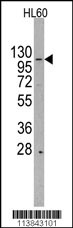
Western blot analysis of anti-HIRA Antibody (Center) (RB13843) in HL60 cell line lysates (35ug/lane). HIRA (arrow) was detected using the purified Pab (1:60 dilution).| Product Name | HIRA Antibody (Center) |
|---|---|
| Antibody Type | Primary Antibodies |
| Antigen Alias | HIRA; DGCR1; HIR; TUPLE1; Protein HIRA; TUP1-like enhancer of split protein 1 |
| Clonality | Polyclonal |
|---|---|
| Isotype | Ig |
| Host Species | Rabbit |
| Tested Applications | WB |
| WB:1:50~100 |
|
| Species Reactivity | Human |
| Concentration | 1mg/ml |
| Gene Synonyms | DGCR1 HIR TUPLE1 |
|---|---|
| Alternative Names | HIRA DGCR1 HIR TUPLE1 Protein HIRA TUP1-like enhancer of split protein 1 |
| Molecular Weight(MW) | 111835 Da |
| Function | Cooperates with ASF1A to promote replication-independent chromatin assembly. Required for the periodic repression of histone gene transcription during the cell cycle. Required for the formation of senescence-associated heterochromatin foci (SAHF) and efficient senescence-associated cell cycle exit |
| Tissue Specificity | Expressed at high levels in kidney, pancreas and skeletal muscle and at lower levels in brain, heart, liver, lung, and placenta |
| Cellular Localization | Nucleus. Nucleus, PML body. Note=Primarily, though not exclusively, localized to the nucleus. Localizes to PML bodies immediately prior to onset of senescence |
| Entrez Gene | 7290 |
|---|
Application
Western blot analysis of anti-HIRA Antibody (Center) (RB13843) in HL60 cell line lysates (35ug/lane). HIRA (arrow) was detected using the purified Pab (1:60 dilution).| Application Notes | WB:1:50~100 |
|---|
| Form | Liquid |
|---|---|
| Storage Instructions | For short-term storage, store at 4° C. For long-term storage, aliquot and store at -20ºC or below. Avoid multiple freeze-thaw cycles. |
| Storage Buffer | Purified polyclonal antibody supplied in PBS with 0.09% (W/V) sodium azide. This antibody is prepared by Saturated Ammonium Sulfate (SAS) precipitation followed by dialysis against PBS. |


