Application
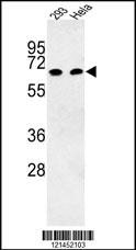
Western blot analysis of HSPA1L Antibody (N-term) in 293, Hela cell line lysates (35ug/lane). HSPA1L (arrow) was detected using the purified Pab.Application
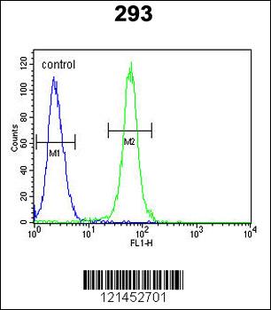
HSPA1L Antibody (N-term) flow cytometric analysis of 293 cells (right histogram) compared to a negative control cell (left histogram).FITC-conjugated goat-anti-rabbit secondary antibodies were used for the analysis.Application
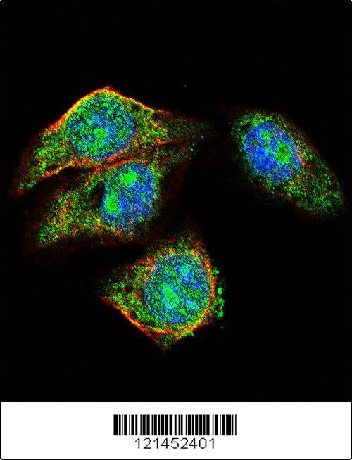
Confocal immunofluorescent analysis of HSPA1L Antibody (N-term)(Cat#AP8798a) with hela cell followed by Alexa Fluor 488-conjugated goat anti-rabbit lgG (green). Actin filaments have been labeled with Alexa Fluor? 555 phalloidin (red). DAPI was used to stain the cell nuclear (blue).| Product Name | HSPA1L Antibody (N-term) |
|---|---|
| Antibody Type | Primary Antibodies |
| Antigen Alias | HSPA1L; Heat shock 70 kDa protein 1-like; Heat shock 70 kDa protein 1-Hom |
| Clonality | Polyclonal |
|---|---|
| Isotype | Ig |
| Host Species | Rabbit |
| Tested Applications | WBFCIF |
| WB:1:100~500 FC |
|
| Species Reactivity | Human |
| Concentration | 1mg/ml |
| Alternative Names | HSPA1L Heat shock 70 kDa protein 1-like Heat shock 70 kDa protein 1-Hom |
|---|---|
| Molecular Weight(MW) | 70375 Da |
| Function | In cooperation with other chaperones, Hsp70s stabilize preexistent proteins against aggregation and mediate the folding of newly translated polypeptides in the cytosol as well as within organelles. These chaperones participate in all these processes through their ability to recognize nonnative conformations of other proteins. They bind extended peptide segments with a net hydrophobic character exposed by polypeptides during translation and membrane translocation, or following stress-induced damage (By similarity) |
| Tissue Specificity | Expressed in spermatids. |
| Entrez Gene | 3305 |
|---|
Application
Western blot analysis of HSPA1L Antibody (N-term) in 293, Hela cell line lysates (35ug/lane). HSPA1L (arrow) was detected using the purified Pab.
Application
HSPA1L Antibody (N-term) flow cytometric analysis of 293 cells (right histogram) compared to a negative control cell (left histogram).FITC-conjugated goat-anti-rabbit secondary antibodies were used for the analysis.
Application
Confocal immunofluorescent analysis of HSPA1L Antibody (N-term)(Cat#AP8798a) with hela cell followed by Alexa Fluor 488-conjugated goat anti-rabbit lgG (green). Actin filaments have been labeled with Alexa Fluor? 555 phalloidin (red). DAPI was used to stain the cell nuclear (blue).| Application Notes | WB:1:100~500 FC |
|---|
| Form | Liquid |
|---|---|
| Storage Instructions | For short-term storage, store at 4° C. For long-term storage, aliquot and store at -20ºC or below. Avoid multiple freeze-thaw cycles. |
| Storage Buffer | Purified polyclonal antibody supplied in PBS with 0.09% (W/V) sodium azide. This antibody is purified through a protein A column, followed by peptide affinity purification. |


