Application
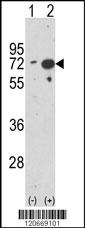
Western blot analysis of HSPA1A (arrow) using rabbit polyclonal HSPA1A Antibody (Center) . 293 cell lysates (2 ug/lane) either nontransfected (Lane 1) or transiently transfected with the HSPA1A gene (Lane 2) .Application
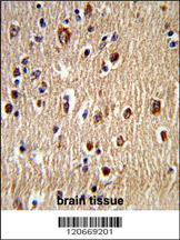
Formalin-fixed and paraffin-embedded human brain tissue reacted with HSPA1A Antibody (Center), which was peroxidase-conjugated to the secondary antibody, followed by DAB staining. This data demonstrates the use of this antibody for immunohistochemistry; clinical relevance has not been evaluated.Application
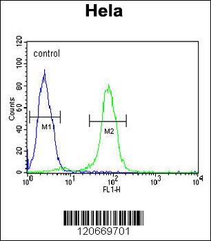
HSPA1A Antibody (Center) flow cytometric analysis of Hela cells (right histogram) compared to a negative control cell (left histogram).FITC-conjugated goat-anti-rabbit secondary antibodies were used for the analysis.Application
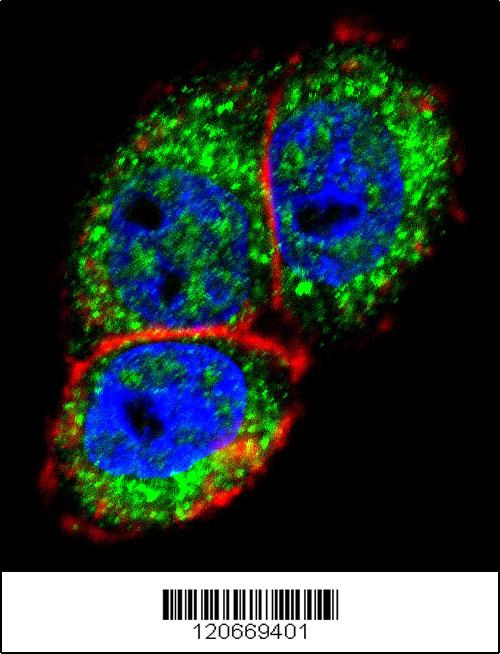
Confocal immunofluorescent analysis of HSPA1A Antibody (Center)(Cat#AP7574d) with Hela cell followed by Alexa Fluor 488-conjugated goat anti-rabbit lgG (green). Actin filaments have been labeled with Alexa Fluor 555 phalloidin (red).DAPI was used to stain the cell nuclear (blue).| Product Name | HSPA1A Antibody (Center) |
|---|---|
| Antibody Type | Primary Antibodies |
| Antigen Alias | HSPA1A; HSPA1; Heat shock 70 kDa protein 1A/1B; Heat shock 70 kDa protein 1/2 |
| Clonality | Polyclonal |
|---|---|
| Isotype | Ig |
| Host Species | Rabbit |
| Tested Applications | WBIHCFCIF |
| WB:1:50~100 IHC |
|
| Species Reactivity | Human |
| Concentration | 1mg/ml |
| Gene Synonyms | HSPA1 |
|---|---|
| Alternative Names | HSPA1A HSPA1 Heat shock 70 kDa protein 1A/1B Heat shock 70 kDa protein 1/2 |
| Molecular Weight(MW) | 70052 Da |
| Function | In cooperation with other chaperones, Hsp70s stabilize preexistent proteins against aggregation and mediate the folding of newly translated polypeptides in the cytosol as well as within organelles. These chaperones participate in all these processes through their ability to recognize nonnative conformations of other proteins. They bind extended peptide segments with a net hydrophobic character exposed by polypeptides during translation and membrane translocation, or following stress-induced damage. In case of rotavirus A infection, serves as a post-attachment receptor for the virus to facilitate entry into the cell |
| Tissue Specificity | HSPA1B is testis-specific. |
| Cellular Localization | Cytoplasm. Note=Localized in cytoplasmic mRNP granules containing untranslated mRNAs |
| Entrez Gene | 3304 |
|---|
Application
Western blot analysis of HSPA1A (arrow) using rabbit polyclonal HSPA1A Antibody (Center) . 293 cell lysates (2 ug/lane) either nontransfected (Lane 1) or transiently transfected with the HSPA1A gene (Lane 2) .
Application
Formalin-fixed and paraffin-embedded human brain tissue reacted with HSPA1A Antibody (Center), which was peroxidase-conjugated to the secondary antibody, followed by DAB staining. This data demonstrates the use of this antibody for immunohistochemistry; clinical relevance has not been evaluated.
Application
HSPA1A Antibody (Center) flow cytometric analysis of Hela cells (right histogram) compared to a negative control cell (left histogram).FITC-conjugated goat-anti-rabbit secondary antibodies were used for the analysis.
Application
Confocal immunofluorescent analysis of HSPA1A Antibody (Center)(Cat#AP7574d) with Hela cell followed by Alexa Fluor 488-conjugated goat anti-rabbit lgG (green). Actin filaments have been labeled with Alexa Fluor 555 phalloidin (red).DAPI was used to stain the cell nuclear (blue).| Application Notes | WB:1:50~100 IHC |
|---|
| Form | Liquid |
|---|---|
| Storage Instructions | For short-term storage, store at 4° C. For long-term storage, aliquot and store at -20ºC or below. Avoid multiple freeze-thaw cycles. |
| Storage Buffer | Purified polyclonal antibody supplied in PBS with 0.09% (W/V) sodium azide. This antibody is prepared by Saturated Ammonium Sulfate (SAS) precipitation followed by dialysis against PBS. |


 Data sheet for OM235465