Application
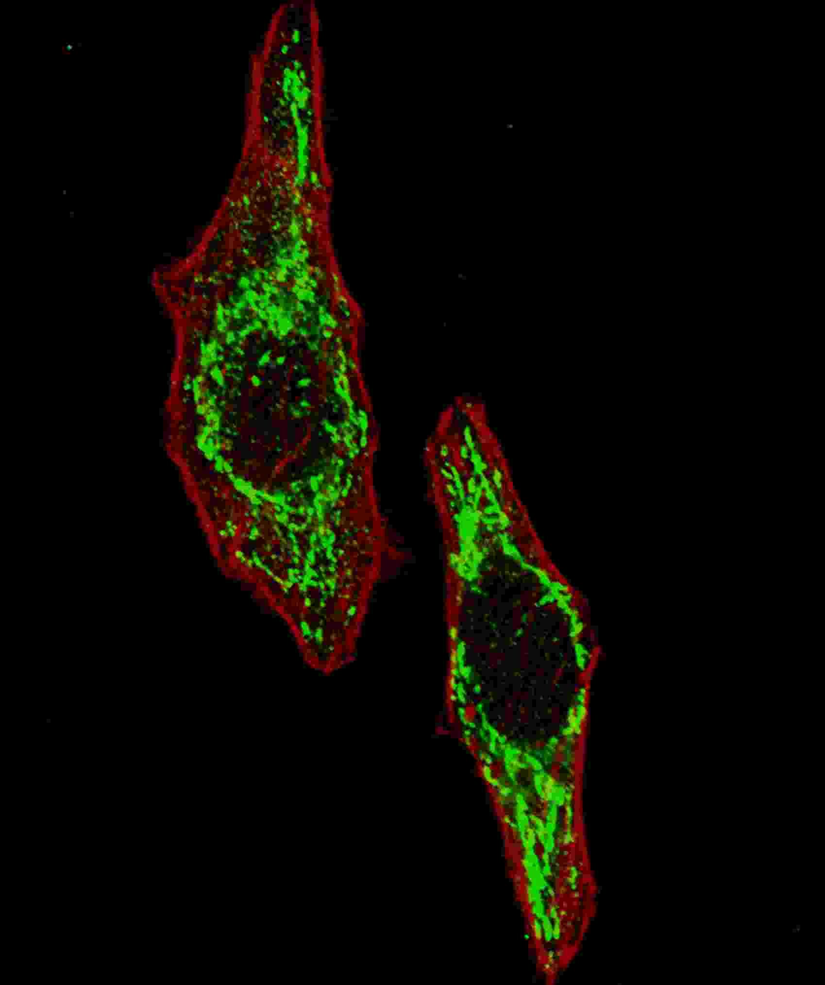
Fluorescent image of U251 cells stained with HSPD1 (Center) antibody. U251 cells were fixed with 4% PFA (20 min), permeabilized with Triton X-100 (0.2%, 30 min). Cells were then incubated with AP2859C HSPD1 (Center) primary antibody (1:100, 2 h at room temperature). For secondary antibody, Alexa Fluor® 488 conjugated donkey anti-rabbit antibody (green) was used (1:1000, 1h). Cytoplasmic actin was counterstained with Alexa Fluor® 555 (red) conjugated Phalloidin (5.25 μM, 25 min). Pictures were taken on a Biorevo microscope (BZ-900, Keyence). Note the highly specific localization of the HSPD1 mainly to the mitochondria, supported by Human Protein Atlas Data (http://www.proteinatlas.org/ENSG00000144381).Application
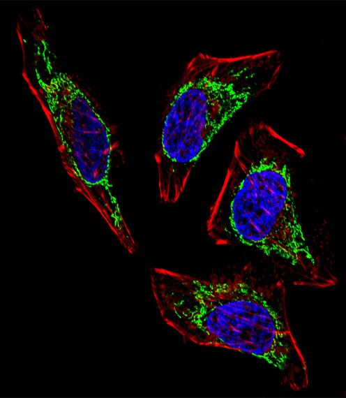
Fluorescent confocal image of Hela cell stained with HSPD1 Antibody (Center)(Cat#AP2859c). Hela cells were fixed with 4% PFA (20 min), permeabilized with Triton X-100 (0.1%, 10 min), then incubated with HSPD1 primary antibody (1:25, 1 h at 37℃). For secondary antibody, Alexa Fluor® 488 conjugated donkey anti-rabbit antibody (green) was used (1:400, 50 min at 37℃).Cytoplasmic actin was counterstained with Alexa Fluor® 555 (red) conjugated Phalloidin (7units/ml, 1 h at 37℃). Nuclei were counterstained with DAPI (blue) (10 µg/ml, 10 min). HSPD1 immunoreactivity is localized to Mitochondria significantly.Application
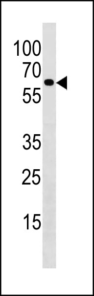
HSPD1 Antibody (Center) (Cat#AP2859c) western blot analysis in A431 cell line lysates (35ug/lane).This demonstrates the HSPD1 antibody detected the HSPD1 protein (arrow).| Product Name | HSPD1 Antibody (Center) |
|---|---|
| Antibody Type | Primary Antibodies |
| Antigen Alias | HSPD1; HSP60; 60 kDa heat shock protein, mitochondrial; 60 kDa chaperonin; Chaperonin 60; Heat shock protein 60; HuCHA60; Mitochondrial matrix protein P1; P60 lymphocyte protein |
| Clonality | Polyclonal |
|---|---|
| Isotype | Ig |
| Host Species | Rabbit |
| Tested Applications | IFWB |
| IF:1:10~50 WB |
|
| Species Reactivity | Human |
| Concentration | 1mg/ml |
| Gene Synonyms | HSP60 |
|---|---|
| Alternative Names | HSPD1 HSP60 60 kDa heat shock protein mitochondrial 60 kDa chaperonin Chaperonin 60 Heat shock protein 60 HuCHA60 Mitochondrial matrix protein P1 P60 lymphocyte protein |
| Molecular Weight(MW) | 61055 Da |
| Function | Implicated in mitochondrial protein import and macromolecular assembly. May facilitate the correct folding of imported proteins. May also prevent misfolding and promote the refolding and proper assembly of unfolded polypeptides generated under stress conditions in the mitochondrial matrix |
| Tissue Specificity | This HSPD1 antibody is generated from rabbits immunized with a KLH conjugated synthetic peptide between 194~223 amino acids from the Center region of human HSPD1. |
| Cellular Localization | Mitochondrion matrix. |
| Entrez Gene | 3329 |
|---|
Application
Fluorescent image of U251 cells stained with HSPD1 (Center) antibody. U251 cells were fixed with 4% PFA (20 min), permeabilized with Triton X-100 (0.2%, 30 min). Cells were then incubated with AP2859C HSPD1 (Center) primary antibody (1:100, 2 h at room temperature). For secondary antibody, Alexa Fluor® 488 conjugated donkey anti-rabbit antibody (green) was used (1:1000, 1h). Cytoplasmic actin was counterstained with Alexa Fluor® 555 (red) conjugated Phalloidin (5.25 μM, 25 min). Pictures were taken on a Biorevo microscope (BZ-900, Keyence). Note the highly specific localization of the HSPD1 mainly to the mitochondria, supported by Human Protein Atlas Data (http://www.proteinatlas.org/ENSG00000144381).
Application
Fluorescent confocal image of Hela cell stained with HSPD1 Antibody (Center)(Cat#AP2859c). Hela cells were fixed with 4% PFA (20 min), permeabilized with Triton X-100 (0.1%, 10 min), then incubated with HSPD1 primary antibody (1:25, 1 h at 37℃). For secondary antibody, Alexa Fluor® 488 conjugated donkey anti-rabbit antibody (green) was used (1:400, 50 min at 37℃).Cytoplasmic actin was counterstained with Alexa Fluor® 555 (red) conjugated Phalloidin (7units/ml, 1 h at 37℃). Nuclei were counterstained with DAPI (blue) (10 µg/ml, 10 min). HSPD1 immunoreactivity is localized to Mitochondria significantly.
Application
HSPD1 Antibody (Center) (Cat#AP2859c) western blot analysis in A431 cell line lysates (35ug/lane).This demonstrates the HSPD1 antibody detected the HSPD1 protein (arrow).| Application Notes | IF:1:10~50 WB |
|---|
| Form | Liquid |
|---|---|
| Storage Instructions | For short-term storage, store at 4° C. For long-term storage, aliquot and store at -20ºC or below. Avoid multiple freeze-thaw cycles. |
| Storage Buffer | Purified polyclonal antibody supplied in PBS with 0.09% (W/V) sodium azide. This antibody is prepared by Saturated Ammonium Sulfate (SAS) precipitation followed by dialysis against PBS. |


