Application
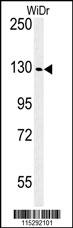
Western blot analysis of IGF1R Antibody (N-term K66) in WiDr cell line lysates (35ug/lane).IGF1R (arrow) was detected using the purified Pab.Application
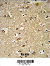
IGF1R Antibody (N-term K66) IHC analysis in formalin fixed and paraffin embedded brain tissue followed by peroxidase conjugation of the secondary antibody and DAB staining. This data demonstrates the use of the IGF1R Antibody (N-term K66) for immunohistochemistry. Clinical relevance has not been evaluated.Application
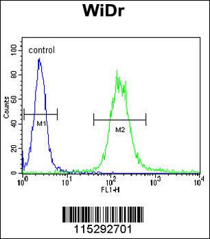
IGF1R Antibody (N-term K66) flow cytometric analysis of WiDr cells (right histogram) compared to a negative control cell (left histogram).FITC-conjugated goat-anti-rabbit secondary antibodies were used for the analysis.Application
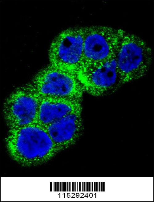
Confocal immunofluorescent analysis of IGF1R Antibody (N-term K66)(Cat#AP7649d) with WiDr cell followed by Alexa Fluor 488-conjugated goat anti-rabbit lgG (green). DAPI was used to stain the cell nuclear (blue).| Product Name | IGF1R Antibody (N-term K66) |
|---|---|
| Antibody Type | Primary Antibodies |
| Antigen Alias | IGF1R; Insulin-like growth factor 1 receptor; Insulin-like growth factor I receptor; CD_antigen=CD221; Insulin-like growth factor 1 receptor alpha chain; Insulin-like growth factor 1 receptor beta chain |
| Clonality | Polyclonal |
|---|---|
| Isotype | Ig |
| Host Species | Rabbit |
| Tested Applications | WBIHCFCIF |
| WB:1:50~100 IHC |
|
| Species Reactivity | Human |
| Concentration | 1mg/ml |
| Alternative Names | IGF1R Insulin-like growth factor 1 receptor Insulin-like growth factor I receptor CD_antigen=CD221 Insulin-like growth factor 1 receptor alpha chain Insulin-like growth factor 1 receptor beta chain |
|---|---|
| Molecular Weight(MW) | 154793 Da |
| Function | Receptor tyrosine kinase which mediates actions of insulin-like growth factor 1 (IGF1). Binds IGF1 with high affinity and IGF2 and insulin (INS) with a lower affinity. The activated IGF1R is involved in cell growth and survival control. IGF1R is crucial for tumor transformation and survival of malignant cell Ligand binding activates the receptor kinase, leading to receptor autophosphorylation, and tyrosines phosphorylation of multiple substrates, that function as signaling adapter proteins including, the insulin-receptor substrates (IRS1/2), Shc and 14-3-3 proteins Phosphorylation of IRSs proteins lead to the activation of two main signaling pathways: the PI3K-AKT/PKB pathway and the Ras-MAPK pathway. The result of activating the MAPK pathway is increased cellular proliferation, whereas activating the PI3K pathway inhibits apoptosis and stimulates protein synthesis Phosphorylated IRS1 can activate the 85 kDa regulatory subunit of PI3K (PIK3R1), leading to activation of several downstream substrates, including protein AKT/PKB. AKT phosphorylation, in turn, enhances protein synthesis through mTOR activation and triggers the antiapoptotic effects of IGFIR through phosphorylation and inactivation of BAD. In parallel to PI3K- driven signaling, recruitment of Grb2/SOS by phosphorylated IRS1 or Shc leads to recruitment of Ras and activation of the ras-MAPK pathway. In addition to these two main signaling pathways IGF1R signals also through the Janus kinase/signal transducer and activator of transcription pathway (JAK/STAT). Phosphorylation of JAK proteins can lead to phosphorylation/activation of signal transducers and activators of transcription (STAT) proteins. In particular activation of STAT3, may be essential for the transforming activity of IGF1R. The JAK/STAT pathway activates gene transcription and may be responsible for the transforming activity. JNK kinases can also be activated by the IGF1R. IGF1 exerts inhibiting activities on JNK activation via phosphorylation and inhibition of MAP3K5/ASK1, which is able to directly associate with the IGF1R |
| Tissue Specificity | Found as a hybrid receptor with INSR in muscle, heart, kidney, adipose tissue, skeletal muscle, hepatoma, fibroblasts, spleen and placenta (at protein level). Expressed in a variety of tissues. Overexpressed in tumors, including melanomas, cancers of the colon, pancreas prostate and kidney |
| Cellular Localization | Membrane; Single-pass type I membrane protein |
| Entrez Gene | 3480 |
|---|
Application
Western blot analysis of IGF1R Antibody (N-term K66) in WiDr cell line lysates (35ug/lane).IGF1R (arrow) was detected using the purified Pab.
Application
IGF1R Antibody (N-term K66) IHC analysis in formalin fixed and paraffin embedded brain tissue followed by peroxidase conjugation of the secondary antibody and DAB staining. This data demonstrates the use of the IGF1R Antibody (N-term K66) for immunohistochemistry. Clinical relevance has not been evaluated.
Application
IGF1R Antibody (N-term K66) flow cytometric analysis of WiDr cells (right histogram) compared to a negative control cell (left histogram).FITC-conjugated goat-anti-rabbit secondary antibodies were used for the analysis.
Application
Confocal immunofluorescent analysis of IGF1R Antibody (N-term K66)(Cat#AP7649d) with WiDr cell followed by Alexa Fluor 488-conjugated goat anti-rabbit lgG (green). DAPI was used to stain the cell nuclear (blue).| Application Notes | WB:1:50~100 IHC |
|---|
| Form | Liquid |
|---|---|
| Storage Instructions | For short-term storage, store at 4° C. For long-term storage, aliquot and store at -20ºC or below. Avoid multiple freeze-thaw cycles. |
| Storage Buffer | Purified polyclonal antibody supplied in PBS with 0.09% (W/V) sodium azide. This antibody is prepared by Saturated Ammonium Sulfate (SAS) precipitation followed by dialysis against PBS. |


