Application
Fluorescent confocal image of HeLa cells stained with ITGA6 (isoform 2 S1064) antibody. HeLa cells were fixed with 4% PFA (20 min), permeabilized with Triton X-100 (0.2%, 30 min). Cells were then incubated with AP2753a ITGA6 (isoform 2 S1064) primary antibody (1:200, 2 h at room temperature). For secondary antibody, Alexa Fluor® 488 conjugated donkey anti-rabbit antibody (green) was used (1:1000, 1h). Nuclei were counterstained with Hoechst 33342 (blue) (10 μg/ml, 5 min). Note the highly specific localization of the ITGA6 mainly to the nucleus, supported by Human Protein Atlas Data (http://www.proteinatlas.org/ENSG00000091409).Application
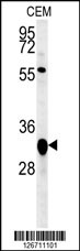
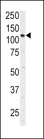
Western blot analysis of anti-ITGA6 Antibody (isoform 2 S1064) in 293 cell line lysates (35ug/lane). ITGA6(arrow) was detected using the purified Pab.Application
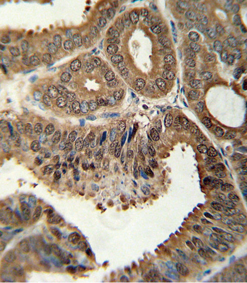
Formalin-fixed and paraffin-embedded human colon carcinoma tissue reacted with ITGA6 Antibody (isoform 2 S1064) , which was peroxidase-conjugated to the secondary antibody, followed by DAB staining. This data demonstrates the use of this antibody for immunohistochemistry; clinical relevance has not been evaluated.| Product Name | ITGA6 Antibody (isoform 2 S1064) |
|---|---|
| Antibody Type | Primary Antibodies |
| Antigen Alias | ITGA6; Integrin alpha-6; CD49 antigen-like family member F; VLA-6; CD_antigen=CD49f; Integrin alpha-6 heavy chain; Integrin alpha-6 light chain |
| Clonality | Polyclonal |
|---|---|
| Isotype | Ig |
| Host Species | Rabbit |
| Tested Applications | IFWBIHC |
| IF:1:200 WB |
|
| Species Reactivity | Human |
| Concentration | 1mg/ml |
| Alternative Names | ITGA6 Integrin alpha-6 CD49 antigen-like family member F VLA-6 CD_antigen=CD49f Integrin alpha-6 heavy chain Integrin alpha-6 light chain |
|---|---|
| Molecular Weight(MW) | 126633 Da |
| Function | Integrin alpha-6/beta-1 is a receptor for laminin on platelets. Integrin alpha-6/beta-4 is a receptor for laminin in epithelial cells and it plays a critical structural role in the hemidesmosome |
| Tissue Specificity | Integrin alpha-6/beta-4 is predominantly expressed by epithelia. Isoforms containing segment X1 are ubiquitously expressed. Isoforms containing segment X1X2 are expressed in heart, kidney, placenta, colon, duodenum, myoblasts and myotubes, and in a limited number of cell lines; they are always coexpressed with the ubiquitous isoform containing segment X1. In some tissues (e.g. Salivary gland), isoforms containing cytoplasmic segment A and isoforms containing segment B are detected while in others, only isoforms containing one cytoplasmic segment are found (segment A in epidermis and segment B in kidney) |
| Cellular Localization | Cell membrane; Single-pass type I membrane protein |
| Entrez Gene | 3655 |
|---|
Application
Fluorescent confocal image of HeLa cells stained with ITGA6 (isoform 2 S1064) antibody. HeLa cells were fixed with 4% PFA (20 min), permeabilized with Triton X-100 (0.2%, 30 min). Cells were then incubated with AP2753a ITGA6 (isoform 2 S1064) primary antibody (1:200, 2 h at room temperature). For secondary antibody, Alexa Fluor® 488 conjugated donkey anti-rabbit antibody (green) was used (1:1000, 1h). Nuclei were counterstained with Hoechst 33342 (blue) (10 μg/ml, 5 min). Note the highly specific localization of the ITGA6 mainly to the nucleus, supported by Human Protein Atlas Data (http://www.proteinatlas.org/ENSG00000091409).
Application
Western blot analysis of anti-ITGA6 Antibody (isoform 2 S1064) in 293 cell line lysates (35ug/lane). ITGA6(arrow) was detected using the purified Pab.
Application
Formalin-fixed and paraffin-embedded human colon carcinoma tissue reacted with ITGA6 Antibody (isoform 2 S1064) , which was peroxidase-conjugated to the secondary antibody, followed by DAB staining. This data demonstrates the use of this antibody for immunohistochemistry; clinical relevance has not been evaluated.| Application Notes | IF:1:200 WB |
|---|
| Form | Liquid |
|---|---|
| Storage Instructions | For short-term storage, store at 4° C. For long-term storage, aliquot and store at -20ºC or below. Avoid multiple freeze-thaw cycles. |
| Storage Buffer | Purified polyclonal antibody supplied in PBS with 0.09% (W/V) sodium azide. This antibody is purified through a protein A column, followed by peptide affinity purification. |


