Application
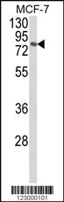
Western blot analysis of JIP1 Antibody (C-term) in MCF-7 cell line lysates (35ug/lane). JIP1 (arrow) was detected using the purified Pab.Application
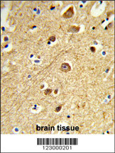
Formalin-fixed and paraffin-embedded human brain tissue reacted with JIP1 Antibody (C-term), which was peroxidase-conjugated to the secondary antibody, followed by DAB staining. This data demonstrates the use of this antibody for immunohistochemistry; clinical relevance has not been evaluated.Application
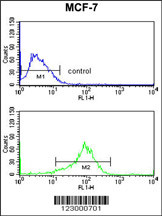
JIP1 Antibody (C-term) flow cytometry analysis of MCF-7 cells (bottom histogram) compared to a negative control cell (top histogram).FITC-conjugated goat-anti-rabbit secondary antibodies were used for the analysis.Application
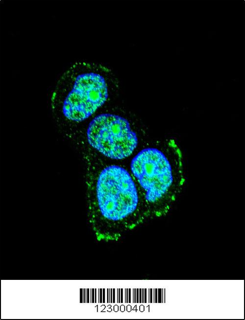
Confocal immunofluorescent analysis of JIP1 Antibody (C-term)(Cat#AP9014b) with MCF-7 cell followed by Alexa Fluor® 488-conjugated goat anti-rabbit lgG (green). DAPI was used to stain the cell nuclear (blue).| Product Name | JIP1 Antibody (C-term) |
|---|---|
| Antibody Type | Primary Antibodies |
| Antigen Alias | MAPK8IP1; IB1; JIP1; PRKM8IP; C-Jun-amino-terminal kinase-interacting protein 1; Islet-brain 1; JNK MAP kinase scaffold protein 1; Mitogen-activated protein kinase 8-interacting protein 1 |
| Clonality | Polyclonal |
|---|---|
| Isotype | Ig |
| Host Species | Rabbit |
| Tested Applications | WBIHCFCIF |
| WB:1:100~500 IHC |
|
| Species Reactivity | Human |
| Concentration | 1mg/ml |
| Gene Synonyms | IB1 JIP1 PRKM8IP |
|---|---|
| Alternative Names | MAPK8IP1 IB1 JIP1 PRKM8IP C-Jun-amino-terminal kinase-interacting protein 1 Islet-brain 1 JNK MAP kinase scaffold protein 1 Mitogen-activated protein kinase 8-interacting protein 1 |
| Molecular Weight(MW) | 77524 Da |
| Function | The JNK-interacting protein (JIP) group of scaffold proteins selectively mediates JNK signaling by aggregating specific components of the MAPK cascade to form a functional JNK signaling module. Required for JNK activation in response to excitotoxic stress. Cytoplasmic MAPK8IP1 causes inhibition of JNK- regulated activity by retaining JNK in the cytoplasm and inhibiting JNK phosphorylation of c-Jun. May also participate in ApoER2-specific reelin signaling. Directly, or indirectly, regulates GLUT2 gene expression and beta-cell function. Appears to have a role in cell signaling in mature and developing nerve terminals. May function as a regulator of vesicle transport, through interactions with the JNK-signaling components and motor proteins (By similarity). Functions as an anti-apoptotic protein and whose level seems to influence the beta-cell death or survival response |
| Tissue Specificity | Highly expressed in brain. Expressed in neurons, localizing to neurite tips in differentiating cells. Also expressed in the pancreas, testis and prostate. Low levels in heart, ovary and small intestine. Decreased levels in pancreatic beta cells sensitize cells to IL-1-beta-induced apoptosis |
| Cellular Localization | Cytoplasm (By similarity). Cytoplasm, perinuclear region (By similarity). Nucleus (By similarity) Endoplasmic reticulum membrane. Mitochondrion membrane Note=Accumulates in cell surface projections. Under certain stress conditions, translocates to the perinuclear region of neurons. In insulin-secreting cells, detected in both the cytoplasm and nucleus (By similarity) |
| Entrez Gene | 9479 |
|---|
Application
Western blot analysis of JIP1 Antibody (C-term) in MCF-7 cell line lysates (35ug/lane). JIP1 (arrow) was detected using the purified Pab.
Application
Formalin-fixed and paraffin-embedded human brain tissue reacted with JIP1 Antibody (C-term), which was peroxidase-conjugated to the secondary antibody, followed by DAB staining. This data demonstrates the use of this antibody for immunohistochemistry; clinical relevance has not been evaluated.
Application
JIP1 Antibody (C-term) flow cytometry analysis of MCF-7 cells (bottom histogram) compared to a negative control cell (top histogram).FITC-conjugated goat-anti-rabbit secondary antibodies were used for the analysis.
Application
Confocal immunofluorescent analysis of JIP1 Antibody (C-term)(Cat#AP9014b) with MCF-7 cell followed by Alexa Fluor® 488-conjugated goat anti-rabbit lgG (green). DAPI was used to stain the cell nuclear (blue).| Application Notes | WB:1:100~500 IHC |
|---|
| Form | Liquid |
|---|---|
| Storage Instructions | For short-term storage, store at 4° C. For long-term storage, aliquot and store at -20ºC or below. Avoid multiple freeze-thaw cycles. |
| Storage Buffer | Purified polyclonal antibody supplied in PBS with 0.09% (W/V) sodium azide. This antibody is purified through a protein A column, followed by peptide affinity purification. |


