Application
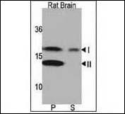
Western blot analysis of anti-LC3 (APG8a) Pab in rat brain lysate. Both lipidated (arrow, II) and non-lipidated APG8a (arrow, I) were detected in membrane fraction (P) but only non-lipidated LC3 was detected in soluble fraction (S).Application
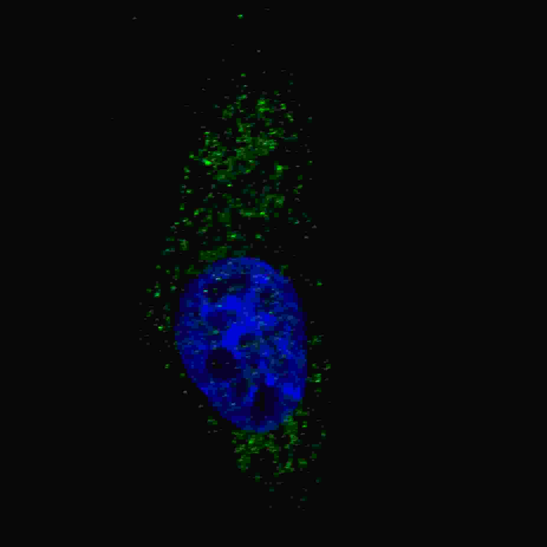
Fluorescent image of U251 cells stained with AP1801a LC3 (APG8A) (N-term) antibody. U251 cells were treated with Chloroquine (50 μM,16h), then fixed with 4% PFA (20 min), permeabilized with Triton X-100 (0.2%, 30 min). Cells were then incubated with AP1801a LC3 (APG8A) (N-term) primary antibody (1:200, 2 h at room temperature). For secondary antibody, Alexa Fluor® 488 conjugated donkey anti-rabbit antibody (green) was used (1:1000, 1h). Nuclei were counterstained with Hoechst 33342 (blue) (10 μg/ml, 5 min). LC3 immunoreactivity is localized to autophagic vacuoles in the cytoplasm of U251 cells.Application
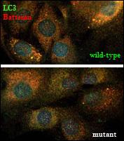
LC3 and the CLN3-encoded protein, battenin, co-immunostaining. Wild-typeand mutant cerebellar cells were costained with #AP1801a, anti-LC3 (APG8a) Abgent antibody (Red)and a monoclonal anti-battenin antibody(Green) and images werecaptured by confocal microscopy. Note the numerous larger vesicular structures, typical ofmature AVs, stained with both the LC3 and battenin antibodies in wild-type cells (arrows),which are rare in mutant cells. Blue=DAPI. Protocol and data courtesy of Dr. Susan Cotman, Center for Human Genetic Research, Massachusetts General Hospital.Application
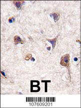
Formalin-fixed and paraffin-embedded human brain tissue reacted with Autophagy LC3 Antibody (APG8a) (N-term), which was peroxidase-conjugated to the secondary antibody, followed by DAB staining. This data demonstrates the use of this antibody for immunohistochemistry; clinical relevance has not been evaluated.Application
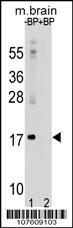
APG8a (MAP1LC3A) Antibody (M1) western blot analysis in mouse brain tissue lysates (35ug/lane).This demonstrates the APG8a (MAP1LC3A) antibody detected the APG8a (MAP1LC3A) protein (arrow).| Product Name | LC3 Antibody (APG8A) (N-term) |
|---|---|
| Antibody Type | Primary Antibodies |
| Antigen Alias | MAP1LC3A; Microtubule-associated proteins 1A/1B light chain 3A; Autophagy-related protein LC3 A; Autophagy-related ubiquitin-like modifier LC3 A; MAP1 light chain 3-like protein 1; MAP1A/MAP1B light chain 3 A; Microtubule-associated protein 1 light chain 3 |
| Clonality | Polyclonal |
|---|---|
| Isotype | Ig |
| Host Species | Rabbit |
| Tested Applications | WBIFIHC |
| WB:1:100~500 IF |
|
| Species Reactivity | Human |
| Concentration | 1mg/ml |
| Alternative Names | MAP1LC3A Microtubule-associated proteins 1A/1B light chain 3A Autophagy-related protein LC3 A Autophagy-related ubiquitin-like modifier LC3 A MAP1 light chain 3-like protein 1 MAP1A/MAP1B light chain 3 A Microtubule-associated protein 1 light chain 3 |
|---|---|
| Molecular Weight(MW) | 14272 Da |
| Function | Involved in formation of autophagosomal vacuoles (autophagosomes) |
| Tissue Specificity | Most abundant in heart, brain, liver, skeletal muscle and testis but absent in thymus and peripheral blood leukocytes |
| Cellular Localization | Cytoplasm, cytoskeleton. Endomembrane system; Lipid-anchor. Cytoplasmic vesicle, autophagosome membrane; Lipid-anchor. Cytoplasmic vesicle, autophagosome. Note=LC3-II binds to the autophagic membranes |
| Entrez Gene | 84557 |
|---|
Application
Western blot analysis of anti-LC3 (APG8a) Pab in rat brain lysate. Both lipidated (arrow, II) and non-lipidated APG8a (arrow, I) were detected in membrane fraction (P) but only non-lipidated LC3 was detected in soluble fraction (S).
Application
Fluorescent image of U251 cells stained with AP1801a LC3 (APG8A) (N-term) antibody. U251 cells were treated with Chloroquine (50 μM,16h), then fixed with 4% PFA (20 min), permeabilized with Triton X-100 (0.2%, 30 min). Cells were then incubated with AP1801a LC3 (APG8A) (N-term) primary antibody (1:200, 2 h at room temperature). For secondary antibody, Alexa Fluor® 488 conjugated donkey anti-rabbit antibody (green) was used (1:1000, 1h). Nuclei were counterstained with Hoechst 33342 (blue) (10 μg/ml, 5 min). LC3 immunoreactivity is localized to autophagic vacuoles in the cytoplasm of U251 cells.
Application
LC3 and the CLN3-encoded protein, battenin, co-immunostaining. Wild-typeand mutant cerebellar cells were costained with #AP1801a, anti-LC3 (APG8a) Abgent antibody (Red)and a monoclonal anti-battenin antibody(Green) and images werecaptured by confocal microscopy. Note the numerous larger vesicular structures, typical ofmature AVs, stained with both the LC3 and battenin antibodies in wild-type cells (arrows),which are rare in mutant cells. Blue=DAPI. Protocol and data courtesy of Dr. Susan Cotman, Center for Human Genetic Research, Massachusetts General Hospital.
Application
Formalin-fixed and paraffin-embedded human brain tissue reacted with Autophagy LC3 Antibody (APG8a) (N-term), which was peroxidase-conjugated to the secondary antibody, followed by DAB staining. This data demonstrates the use of this antibody for immunohistochemistry; clinical relevance has not been evaluated.
Application
APG8a (MAP1LC3A) Antibody (M1) western blot analysis in mouse brain tissue lysates (35ug/lane).This demonstrates the APG8a (MAP1LC3A) antibody detected the APG8a (MAP1LC3A) protein (arrow).| Application Notes | WB:1:100~500 IF |
|---|
| Form | Liquid |
|---|---|
| Storage Instructions | For short-term storage, store at 4° C. For long-term storage, aliquot and store at -20ºC or below. Avoid multiple freeze-thaw cycles. |
| Storage Buffer | Purified polyclonal antibody supplied in PBS with 0.09% (W/V) sodium azide. This antibody is prepared by Saturated Ammonium Sulfate (SAS) precipitation followed by dialysis against PBS. |


