Application
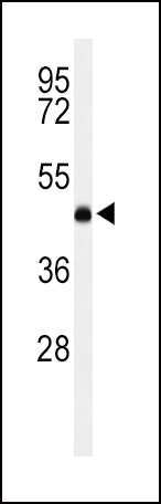
Western blot analysis of LCAT Antibody (Center) in K562 cell line lysates (35ug/lane). LCAT (arrow) was detected using the purified Pab.Application
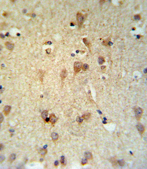
LCAT Antibody (Center) IHC analysis in formalin fixed and paraffin embedded brain tissue followed by peroxidase conjugation of the secondary antibody and DAB staining. This data demonstrates the use of the LCAT Antibody (Center) for immunohistochemistry. Clinical relevance has not been evaluated.Application
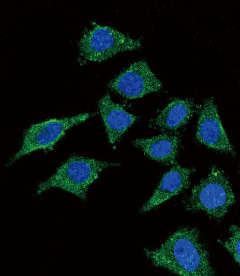
Confocal immunofluorescent analysis of LCAT Antibody (Center) with 293 cell followed by Alexa Fluor搴?488-conjugated goat anti-rabbit lgG (green).DAPI was used to stain the cell nuclear (blue).Application
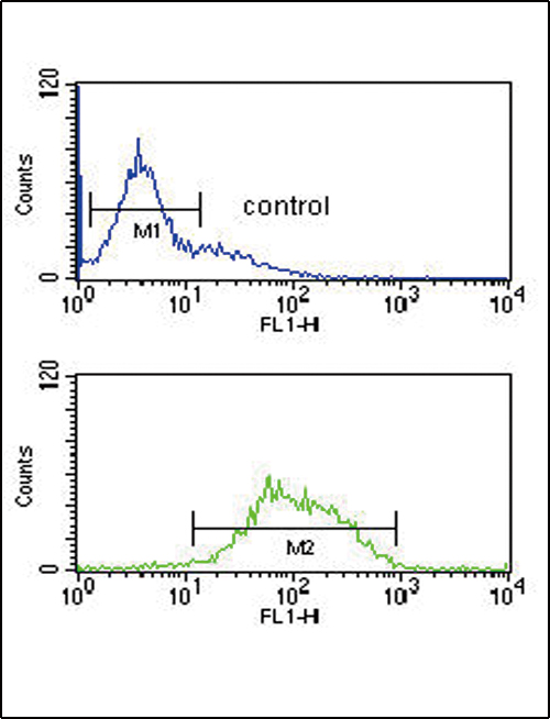
LCAT Antibody (Center) flow cytometric analysis of K562 cells (bottom histogram) compared to a negative control cell (top histogram).FITC-conjugated goat-anti-rabbit secondary antibodies were used for the analysis.| Product Name | LCAT Antibody (Center) |
|---|---|
| Antibody Type | Primary Antibodies |
| Antigen Alias | LCAT; Phosphatidylcholine-sterol acyltransferase; Lecithin-cholesterol acyltransferase; Phospholipid-cholesterol acyltransferase |
| Clonality | Polyclonal |
|---|---|
| Isotype | Ig |
| Host Species | Rabbit |
| Tested Applications | WBIHCIFFC |
| WB:1:100~500 IHC |
|
| Species Reactivity | Human |
| Concentration | 1mg/ml |
| Alternative Names | LCAT Phosphatidylcholine-sterol acyltransferase Lecithin-cholesterol acyltransferase Phospholipid-cholesterol acyltransferase |
|---|---|
| Molecular Weight(MW) | 49578 Da |
| Function | Central enzyme in the extracellular metabolism of plasma lipoproteins. Synthesized mainly in the liver and secreted into plasma where it converts cholesterol and phosphatidylcholines (lecithins) to cholesteryl esters and lysophosphatidylcholines on the surface of high and low density lipoproteins (HDLs and LDLs) The cholesterol ester is then transported back to the liver. Has a preference for plasma 16:0-18:2 or 18:O-18:2 phosphatidylcholines Also produced in the brain by primary astrocytes, and esterifies free cholesterol on nascent APOE-containing lipoproteins secreted from glia and influences cerebral spinal fluid (CSF) APOE- and APOA1 levels. Together with APOE and the cholesterol transporter ABCA1, plays a key role in the maturation of glial-derived, nascent lipoproteins. Required for remodeling high-density lipoprotein particles into their spherical forms |
| Tissue Specificity | Expressed mainly in brain, liver and testes. Secreted into plasma and cerebral spinal fluid. Expressed in Hep- G2 cell line |
| Cellular Localization | Secreted. Note=Secreted into blood plasma. Produced in astrocytes and secreted into cerebral spinal fluid (CSF) |
| Entrez Gene | 3931 |
|---|
Application
Western blot analysis of LCAT Antibody (Center) in K562 cell line lysates (35ug/lane). LCAT (arrow) was detected using the purified Pab.
Application
LCAT Antibody (Center) IHC analysis in formalin fixed and paraffin embedded brain tissue followed by peroxidase conjugation of the secondary antibody and DAB staining. This data demonstrates the use of the LCAT Antibody (Center) for immunohistochemistry. Clinical relevance has not been evaluated.
Application
Confocal immunofluorescent analysis of LCAT Antibody (Center) with 293 cell followed by Alexa Fluor搴?488-conjugated goat anti-rabbit lgG (green).DAPI was used to stain the cell nuclear (blue).
Application
LCAT Antibody (Center) flow cytometric analysis of K562 cells (bottom histogram) compared to a negative control cell (top histogram).FITC-conjugated goat-anti-rabbit secondary antibodies were used for the analysis.| Application Notes | WB:1:100~500 IHC |
|---|
| Form | Liquid |
|---|---|
| Storage Instructions | For short-term storage, store at 4° C. For long-term storage, aliquot and store at -20ºC or below. Avoid multiple freeze-thaw cycles. |
| Storage Buffer | Purified polyclonal antibody supplied in PBS with 0.09% (W/V) sodium azide. This antibody is purified through a protein A column, followed by peptide affinity purification. |


