Application
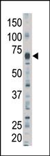
Western blot analysis of anti-LIMK2 Pab in mouse brain tissue lysate. LIMK2 (arrow) was detected using purified Pab. Secondary HRP-anti-rabbit was used for signal visualization with chemiluminescence.| Product Name | LIM Kinase 2 (LIMK2) Antibody (N-term) |
|---|---|
| Antibody Type | Primary Antibodies |
| Antigen Alias | LIMK2; LIM domain kinase 2 |
| Clonality | Polyclonal |
|---|---|
| Isotype | Ig |
| Host Species | Rabbit |
| Tested Applications | WB |
| WB:1:100~500 |
|
| Species Reactivity | Human |
| Concentration | 1mg/ml |
| Alternative Names | LIMK2 LIM domain kinase 2 |
|---|---|
| Molecular Weight(MW) | 72232 Da |
| Function | Displays serine/threonine-specific phosphorylation of myelin basic protein and histone (MBP) in vitro |
| Tissue Specificity | Highest expression in the placenta; moderate level in liver, lung, kidney, and pancreas. LIMK2a is found to be more abundant then LIMK2b in liver, colon, stomach, and spleen, while in brain, kidney, and placenta LIMK2b is the dominant form In adult lung, both LIMK2a and LIMK2b is nearly equally observed |
| Cellular Localization | Isoform LIMK2a: Cytoplasm. Nucleus. Note=Isoform LIMK2a is distributed in the cytoplasm and the nucleus |
| Entrez Gene | 3985 |
|---|---|
| Nucleotide Accession | NP_057952.1 |
Application
Western blot analysis of anti-LIMK2 Pab in mouse brain tissue lysate. LIMK2 (arrow) was detected using purified Pab. Secondary HRP-anti-rabbit was used for signal visualization with chemiluminescence.| Application Notes | WB:1:100~500 |
|---|
| Form | Liquid |
|---|---|
| Storage Instructions | For short-term storage, store at 4° C. For long-term storage, aliquot and store at -20ºC or below. Avoid multiple freeze-thaw cycles. |
| Storage Buffer | Purified polyclonal antibody supplied in PBS with 0.09% (W/V) sodium azide. This antibody is purified through a protein G column, eluted with high and low pH buffers and neutralized immediately, followed by dialysis against PBS. |


