Application
Western blot analysis of anti-LIN28B Pab in K562 cell line lysates (35ug/lane). LIN28B(arrow) was detected using the purified Pab (1:60 dilution).Application
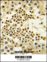
Formalin-fixed and paraffin-embedded human testis tissue reacted with LIN28B antibody (Center) , which was peroxidase-conjugated to the secondary antibody, followed by DAB staining. This data demonstrates the use of this antibody for immunohistochemistry; clinical relevance has not been evaluated.Application
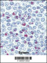
Formalin-fixed and paraffin-embedded human Spleen tissue reacted with LIN28B antibody (Center) , which was peroxidase-conjugated to the secondary antibody, followed by AEC staining. This data demonstrates the use of this antibody for immunohistochemistry; clinical relevance has not been evaluated.Application
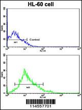
Flow cytometric analysis of HL-60 cells using LIN28B Antibody (Center)(bottom histogram) compared to a negative control cell (top histogram). FITC-conjugated goat-anti-rabbit secondary antibodies were used for the analysis.Application
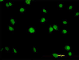
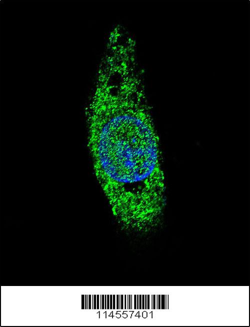
Confocal immunofluorescent analysis of LIN28B Antibody (Center) (Cat#AP1485c) with HepG2 cell followed by Alexa Fluor 488-conjugated goat anti-rabbit lgG (green). DAPI was used to stain the cell nuclear (blue).| Product Name | LIN28B Antibody (Center) |
|---|---|
| Antibody Type | Primary Antibodies |
| Antigen Alias | LIN28B; CSDD2; Protein lin-28 homolog B |
| Clonality | Polyclonal |
|---|---|
| Isotype | Ig |
| Host Species | Rabbit |
| Tested Applications | WBIHCFCIF |
| WB:1:50~100 IHC |
|
| Species Reactivity | Human |
| Concentration | 1mg/ml |
| Gene Synonyms | CSDD2 |
|---|---|
| Alternative Names | LIN28B CSDD2 Protein lin-28 homolog B |
| Molecular Weight(MW) | 27084 Da |
| Function | Acts as a suppressor of microRNA (miRNA) biogenesis by specifically binding the precursor let-7 (pre-let-7), a miRNA precursor. Acts by binding pre-let-7 and recruiting ZCCHC11/TUT4 uridylyltransferase, leading to the terminal uridylation of pre- let-7. Uridylated pre-let-7 miRNAs fail to be processed by Dicer and undergo degradation. Specifically recognizes the 5'-GGAG-3' motif in the terminal loop of pre-let-7. Also recognizes and binds non pre-let-7 pre-miRNAs that contain the 5'-GGAG-3' motif in the terminal loop, leading to their terminal uridylation and subsequent degradation. Mediates MYC-mediated let-7 repression Isoform 1, when overexpressed, stimulates growth of the breast adenocarcinoma cell line MCF-7. Isoform 2 has no effect on cell growth |
| Tissue Specificity | High expression in testis, fetal liver, placenta and in hepatocellular carcinoma (HCC). Isoform 1 is only detected in moderately and poorly differentiated HCC tissues and placenta (at protein level). Isoform 2 is detected in fetal liver, non-tumor liver tissues, as well as well-differentiated tumor tissues (at protein level) |
| Cellular Localization | Cytoplasm. Nucleus. Note=Predominantly cytoplasmic at G1 phase, accumulates in the nucleus in S and G2 phases. The frequency of nuclear localization in S and in G2 phases is 60% and 30%, respectively |
| Entrez Gene | 389421 |
|---|
Application
Western blot analysis of anti-LIN28B Pab in K562 cell line lysates (35ug/lane). LIN28B(arrow) was detected using the purified Pab (1:60 dilution).
Application
Formalin-fixed and paraffin-embedded human testis tissue reacted with LIN28B antibody (Center) , which was peroxidase-conjugated to the secondary antibody, followed by DAB staining. This data demonstrates the use of this antibody for immunohistochemistry; clinical relevance has not been evaluated.
Application
Formalin-fixed and paraffin-embedded human Spleen tissue reacted with LIN28B antibody (Center) , which was peroxidase-conjugated to the secondary antibody, followed by AEC staining. This data demonstrates the use of this antibody for immunohistochemistry; clinical relevance has not been evaluated.
Application
Flow cytometric analysis of HL-60 cells using LIN28B Antibody (Center)(bottom histogram) compared to a negative control cell (top histogram). FITC-conjugated goat-anti-rabbit secondary antibodies were used for the analysis.
Application
Confocal immunofluorescent analysis of LIN28B Antibody (Center) (Cat#AP1485c) with HepG2 cell followed by Alexa Fluor 488-conjugated goat anti-rabbit lgG (green). DAPI was used to stain the cell nuclear (blue).| Application Notes | WB:1:50~100 IHC |
|---|
| Form | Liquid |
|---|---|
| Storage Instructions | For short-term storage, store at 4° C. For long-term storage, aliquot and store at -20ºC or below. Avoid multiple freeze-thaw cycles. |
| Storage Buffer | Purified polyclonal antibody supplied in PBS with 0.09% (W/V) sodium azide. This antibody is prepared by Saturated Ammonium Sulfate (SAS) precipitation followed by dialysis against PBS. |


