Application
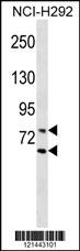
LMNA Antibody (Center) western blot analysis in NCI-H292 cell line lysates (35ug/lane).This demonstrates the LMNA Antibody detected the LMNA protein (arrow).| Product Name | LMNA Antibody (Center) |
|---|---|
| Antibody Type | Primary Antibodies |
| Antigen Alias | LMNA; LMN1; Prelamin-A/C; Lamin-A/C; 70 kDa lamin; Renal carcinoma antigen NY-REN-32 |
| Clonality | Polyclonal |
|---|---|
| Isotype | Ig |
| Host Species | Rabbit |
| Tested Applications | WB |
| WB:1:100~500 |
|
| Species Reactivity | Human |
| Concentration | 1mg/ml |
| Gene Synonyms | LMN1 |
|---|---|
| Alternative Names | LMNA LMN1 Prelamin-A/C Lamin-A/C 70 kDa lamin Renal carcinoma antigen NY-REN-32 |
| Molecular Weight(MW) | 74139 Da |
| Function | Lamins are components of the nuclear lamina, a fibrous layer on the nucleoplasmic side of the inner nuclear membrane, which is thought to provide a framework for the nuclear envelope and may also interact with chromatin. Lamin A and C are present in equal amounts in the lamina of mammals. Plays an important role in nuclear assembly, chromatin organization, nuclear membrane and telomere dynamics |
| Tissue Specificity | In the arteries, prelamin-A/C accumulation is not observed in young healthy vessels but is prevalent in medial vascular smooth muscle cells (VSMCs) from aged individuals and in atherosclerotic lesions, where it often colocalizes with senescent and degenerate VSMCs. Prelamin-A/C expression increases with age and disease. In normal aging, the accumulation of prelamin-A/C is caused in part by the down-regulation of ZMPSTE24/FACE1 in response to oxidative stress |
| Cellular Localization | Nucleus. Nucleus envelope. Note=Farnesylation of prelamin-A/C facilitates nuclear envelope targeting and subsequent cleaveage by ZMPSTE24/FACE1 to remove the farnesyl group produces mature lamin-A/C, which can then be inserted into the nuclear lamina. EMD is required for proper localization of non-farnesylated prelamin-A/C |
| Entrez Gene | 4000 |
|---|---|
| Nucleotide Accession | NP_733821.1 |
Application
LMNA Antibody (Center) western blot analysis in NCI-H292 cell line lysates (35ug/lane).This demonstrates the LMNA Antibody detected the LMNA protein (arrow).| Application Notes | WB:1:100~500 |
|---|
| Form | Liquid |
|---|---|
| Storage Instructions | For short-term storage, store at 4° C. For long-term storage, aliquot and store at -20ºC or below. Avoid multiple freeze-thaw cycles. |
| Storage Buffer | Purified polyclonal antibody supplied in PBS with 0.09% (W/V) sodium azide. This antibody is purified through a protein A column, followed by peptide affinity purification. |


